 |
|
|
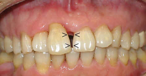
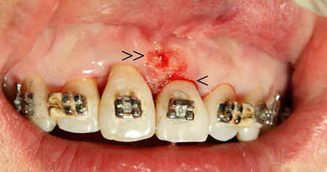
Fig.1 |
Fig.2 |
 |
|
|
Fig.3 |
Fig.4 |
 |
 |
|
Fig.5 |
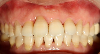
Fig.6 |
 |
 |
|
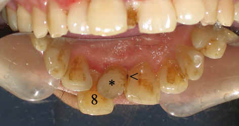
Fig.7 |
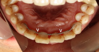
Fig.8 |
|
|
|
Fig.1 |
Fig.2 |
|
|
|
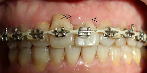
Fig.3 |
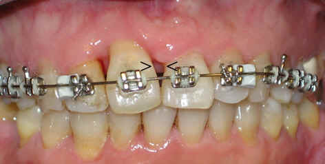
Fig.4 |
|
|
|
Fig.5 |
Fig.6 |
|
|
|
|
Fig.7 |
Fig.8 |
Dental Education Lecture: Progress of Braces
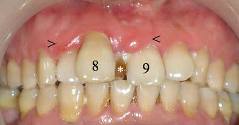
Mr. Huang has an extra tooth (* in Fig.1,7) between two front teeth (8,9) , interfering with local oral hygiene with red and bleeding gums (arrowheads in Fig.1). There are cavities between the extra tooth and the tooth #9 (arrowhead in Fig.7). After removing the tiny tooth and deep cleaning, the oral hygiene improves, but the patient complains that he cannot speak properly, air coming out between the front teeth (arrowheads in Fig.2). After a few months of wearing braces, the space closes partially; the space in the upper portion remains (arrowheads in Fig.3). Adjustment of braces closes the space completely, but there is extra gum tissue between the two front teeth (double arrowheads in Fig.4). The tooth #9 looks too short. We trim off extra tissue under local anesthesia (Fig.5). Finally, Mr. Huang is pleased with the result (Fig.6,8). Fig.7,8 are the inside views of the front teeth through a reflecting mirror. Arrowheads in Fig.8 point to a so-called fixed retainer to keep the teeth in place. There is tendency for the teeth to return to bad position without retainer. We meet this patient in a ballet performance 2.5 years after braces were removed. His teeth look great. We will update his photos when they become available.
For adults who need braces, it is extremely important to have proper gum treatment (such as deep cleaning) before braces. During brace treatment, patients should keep teeth clean. Otherwise, braces make gum disease worse.
Xin Wei, DDS, PhD, MS 1st edition 11/24/2009, last revision 06/23/2012