|
|
|
|
|
|
|
|
|
|
The Earliest Cavities
The earliest cavities can be detected by X-ray, but they may not need treatment immediately. If we keep using floss, the earliest cavities may return to normal.
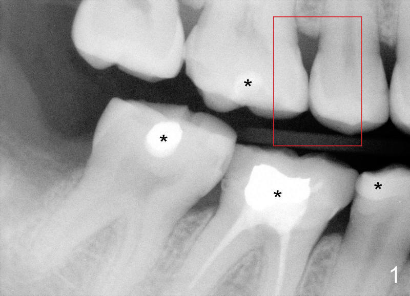
Ms. Li has a lot of fillings (Fig.1 *), suggesting that she has had quite a few of cavities before. Routine X-ray shows that something is abnormal between the teeth (red box).
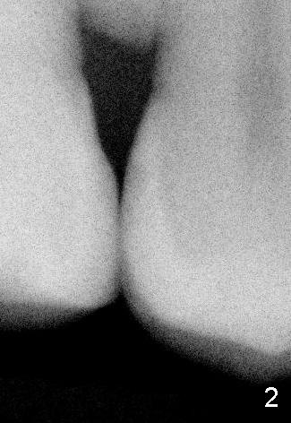
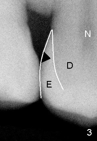
Fig.2 is a blow up picture of the red box. We may not see anything striking. Fig.3 has some labeling added to Fig.2. The white outline is the enamel, the outermost tooth structure (E). Underneath the enamel is the dentin (D). A little further inside is the nerve (N). The enamel, the hardest tissue of our body, is the whitest in X-ray. When it becomes abnormal, such as loss of calcium, it is dark as shown as black triangle in Fig.2,3. The black triangle barely crosses the half thickness of the enamel. It is the earliest cavity.
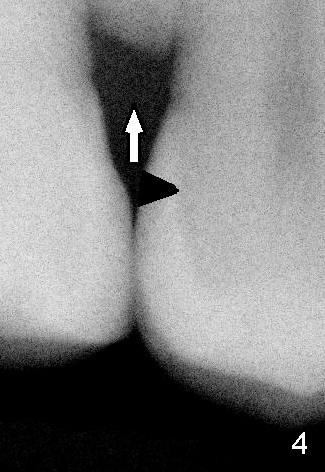
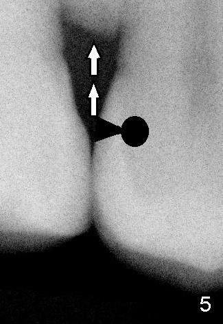
With further loss of calcium (arrow in Fig.4), the cavity becomes deeper, across the whole thickness of the enamel, as compared to Fig.3. With loss of more calcium (double arrow in Fig.5), cavity extends to the dentin (black circle), getting closer to the nerve, as mentioned in Fig.3. Cavities shown in Fig.4,5 need fillings in timely manners.
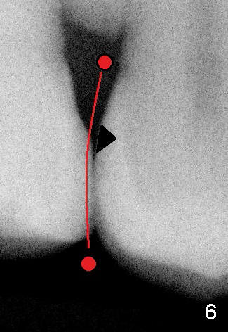
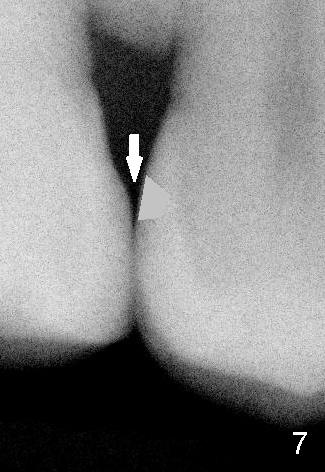
The earliest cavity shown in Fig.3,6 may not need immediate treatment. If floss is used to clean up the area between the teeth (red circle and line in Fig.6), our enamel is getting calcium from saliva(down arrow toward the tooth in Fig.7) instead of losing it. Gradually the cavity may be stopping. The density of the enamel may return to normal (gray triangle in Fig.7). It is self repairing. X-ray should be taken every 6 months to confirm the reverting process.
When cavities are too deep as shown in Fig.4,5, rigorous flossing may not be able to get out bacteria and their food from the deepest portion of the cavities. The cavities continue going deeper. Therefore immediate treatment is necessary.
Xin Wei, DDS, PhD, MS 1st edition 04/19/2012, last revision 04/19/2012