|
|
|
|
| Fig.1 | Fig.2 | Fig.3 |
|
|
|
|
| Fig.4 | Fig.5 | Fig.6 |
|
|
|
|
| Fig.7 | Fig.8 | Fig.9 |
|
|
||
| Fig.10 |
Dental Education Lecture: Drawbacks of Long Bridge
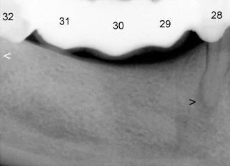
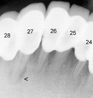
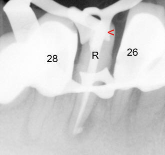
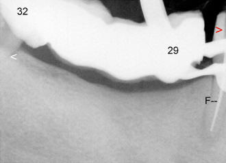
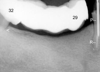
Ms. Zhao has lost two molars (#30,31) and one premolar (#29, Fig.1). In order to make a bridge, several supporting teeth were shaved and capped (#24 to 28 and #32, Fig.1,2). All crowns are connected to form a long bridge. It is extremely difficult to keep good oral hygiene underneath the bridge. One day, the tooth #27 with an over-sized cap has nerve and root tip infection (arrowhead in Fig.2). To facilitate root canal (R in Fig.3), the cap is removed; a hole is made through the natural tooth (red arrowhead). The infection is gone afterward.
But three months later, the neighboring tooth, #28, has similar root tip infection (black arrowhead in Fig.1). Root canal is necessary. It is the best to remove the cap as well. But the patient declines to have the cap taken out, because after taking out the cap of #27, the back portion of the bridge become loose. As expected, removing the cap for #28 change the back portion of the bridge into a cantilever one. The latter will be much looser.
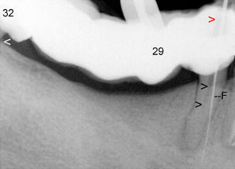
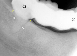
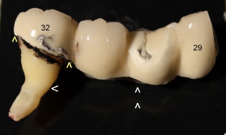
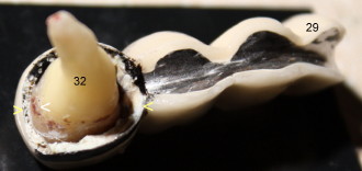
Anyway, root canal is attempted by cutting a hole through the overly-built cap (red arrowhead in Fig.4). A file (F; used to clean the nerve) goes to a wrong place, away from root canal (double arrowheads). After re-obtaining consent from Ms. Zhao, the cap is removed; a file goes to the right canal smoothly (F in Fig.5). Then root canal is finished (R in Fig.6) and a post is placed (P) for the tooth #28. Now the bridge from #29 to #32 belongs to cantilever. It is very loose. The last supporting tooth #32 cannot support the chewing load and has pain sometimes. It has a single skinny root (white arrowheads in Fig.7); the cap is also over-sized with large steps (yellow arrowheads). Food is easily trapped underneath the steps. Finally the bridge and the last supporting tooth (wisdom tooth) are removed (Fig.8). There are tons of nasty stuffs underneath the bridge, especially underneath the cap of #32 (yellow arrowheads). Fig.8 is taken after the tooth is soaked in bleach for 24 hours. Soft junks are gone, but tough materials such as stain and tar remain (Fig.8). Let us take a look at the undersurface of the bridge as seen by double arrowheads in Fig.8. The margins of the cap (yellow arrowheads in Fig.9) does not fit the root of #32 with formation of tartars (white arrowhead).
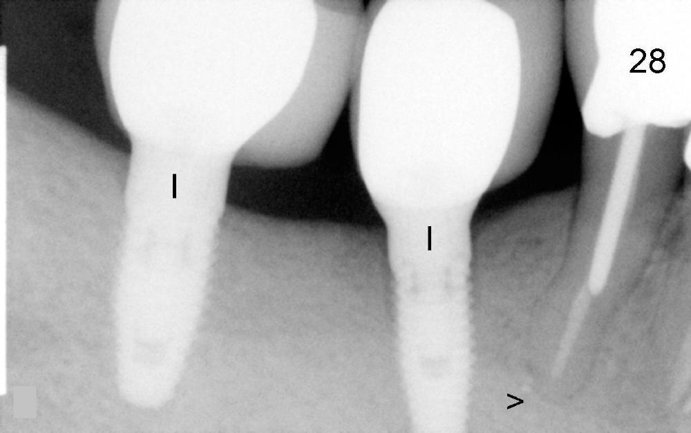
When we lose several teeth, bridge is not a good option. Although it is fixed and easy to chew, it causes many problems in the future as shown above. A denture is another option. It is easy to clean and good for our natural teeth. The best option is placement of several implants. Finally the patient gets two implants in the missing tooth area (Fig.10 I). There is no root tip infection three years after root canal (>, as compared to Fig.4-6).
Xin Wei, DDS, PhD, MS 1st edition 10/27/2010, last revision 01/25/2013