|
|
|
|
|
|
|
|
|
|
Dental Education Lecture: Bone Manipulation for Implant
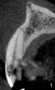
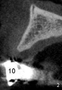
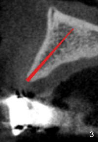
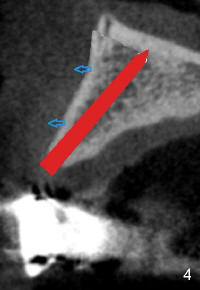
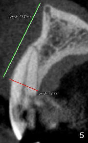
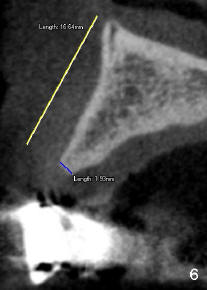
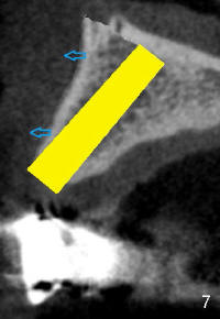
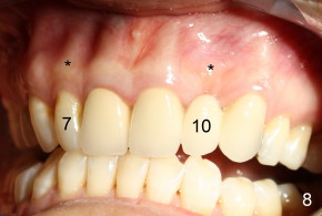
When a tooth such as the upper left lateral incisor (#10 in Fig.8) is extracted, the gums and bone in the root area (*) is shrinking (ditching in), affecting appearance. By contrast, the root of the corresponding tooth (* above #7: upper right lateral incisor) is bulging out, the natural appearance. We are going to make slices through the teeth #7 and 10 and their roots (long axis). We can see that bone is shrinking after tooth is extracted (Fig.2,6 vs. Fig.1,5), particularly the width: from 7.21 mm to 1.93 mm (compare Fig.5,6). To place an implant, we need to increase the width of the bone. There are two ways to do it. The first one is to do bone graft, i.e., placing a piece of bone over the shrinking bone, making the latter thicker. The second way is to expand the bone. Contrast to our thought, our bone is malleable. It can be expanded like a balloon. But the surgeon needs to be careful, avoiding the balloon to pop. Instead of using air, the surgeon uses a set of chisels to expand the bone. First, a very thin chisel (like a thin blade) (red in Fig.3) is inserted into the middle of the bone. Gradually, thicker and thicker chisels (red in Fig.4) are used to make the outside layer of the bone move outward (arrows). Finally, enough space is created to hold a proper sized implant (yellow in Fig.7). With further outward movement of the outside layer of the bone (arrows), the width of the bone is almost equal to that in Fig.1 or 5. The appearance must also improve. In fact, bone does not have nerve. With proper local anesthesia, bone expansion is pain free.
Xin Wei, DDS, PhD, MS 1st edition 03/15/2011, last revision 09/27/2012