|
|
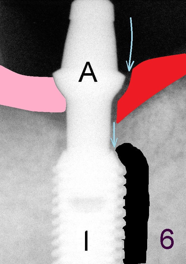
Fig.6 is a schematic drawing to show how the two types of the gums glue to implant/abutment (in the same way for natural teeth). On the left hand side, the tough gums (pink area) firmly glue to the abutment to form a tight seal so that bacteria cannot go in. There is no infection around the implant (I) underneath. On the right hand side of Fig.6, the loose gums (red area) are not able to stick to the surface of the abutment tightly. There is a gap between the loose gums and the abutment. The gap is big enough for tinny tiny germs to enter (long blue arrow), travel all the way down to the implant (short blue arrow) and cause severe infection in the bone around the implant. The end result is loss of bone around the implant (black area). The implant may be loose. This disease is called peri-implantitis. Return to main article
Xin Wei, DDS, PhD, MS 1st edition 08/21/2012, last revision 09/27/2012