 |
 |
 |
|
Fig.1
|
Fig.2
|
Fig.3
|
 |

|
 |
|
Fig.4
|
Fig.5
|
Fig.6
|
 |
 |
 |
|
Fig.7
|
Fig.8
|
Fig.9
|
 |
 |
|
|
Fig.10 |
Fig.11 |
|
Dental Education Lecture: Model Surgery for Implant
To save one minute in surgery, implant surgeons have to spend ten or more
minutes to prepare for it. The best way to prepare for implant surgery is
to do model surgery and make a surgical guide.
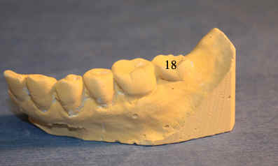
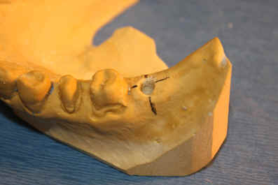
Mrs. Lee agrees to remove a cantilever
bridge to place an implant for the missing tooth #18 (Fig.1).
Impression is taken before and after bridge removal to make models as shown in
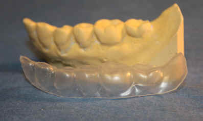
Fig.1 and 4, respectively. The first model is used to make a plastic stent
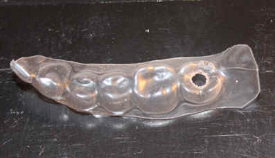
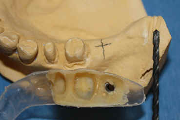
(Fig.2). A hole is made in the middle of the missing tooth (Fig.3).
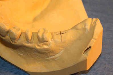
The hole is approximately the center of implant placement. Another way to
determine the point of the future implant placement is to draw a cross
line on the 2nd model (Fig.4). To double check the accuracy of implant
model surgery, the stent with the hole in Fig.3 is to be placed on the model in
Fig.4. It appears that the two ways of determination of the center of
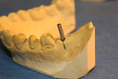
implant placement match well (Fig.5). After removal of the stent, a hole
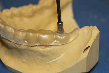
is drilled on the model (Fig.6). Then a metal rod is inserted into the
hole to see whether the orientation of implant placement is right or not
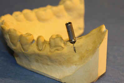
(Fig.7). If the orientation is ok, a metal tube slides over the rod
(Fig.8). Acrylic (plastic) is used to fixate the tube in the stent
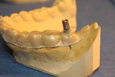
(Fig.9). Let us flip the stent with the tube (i.e., surgical guide;
without the rod) over and place it next to a new model (Fig.10). The
latter in fact will be our patient (Mrs. Lee). When the stent is flipped
back on the model, a real implant surgery can be started (Fig.11).
The preparation on the model is tedious, but it provides precision for the
surgery. Good plan gets half done. The patient will recover quick
from the
procedure.
Xin Wei, DDS, PhD, MS 1st edition 12/08/2009, last revision
09/28/2012