|
|
|
|
|
|
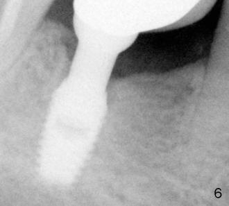
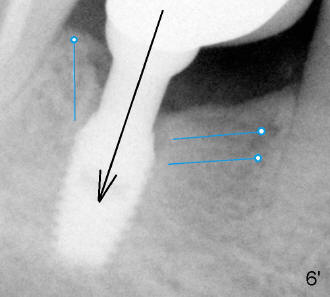
Fig.6 A year later, bone continues to grow denser (whiter in x-ray) all around the implant. The bone has distinct patterns. Tiny bony structures (trabeculae) (Fig.6 ' (Fig.6 with illustrations) blue lines) appear to be inserted obliquely or perpendicularly into the surface of the implant. Under normal chewing (arrow), the implant (like a suspension bed) stretch these trabeculae (ropes attached to the bed), making them stronger and stronger (denser and denser). Three blue circles are equivalent to nails driven into the tree trunks on the either side of the bed.
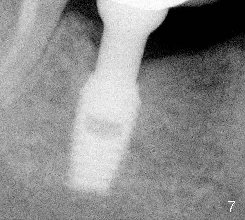
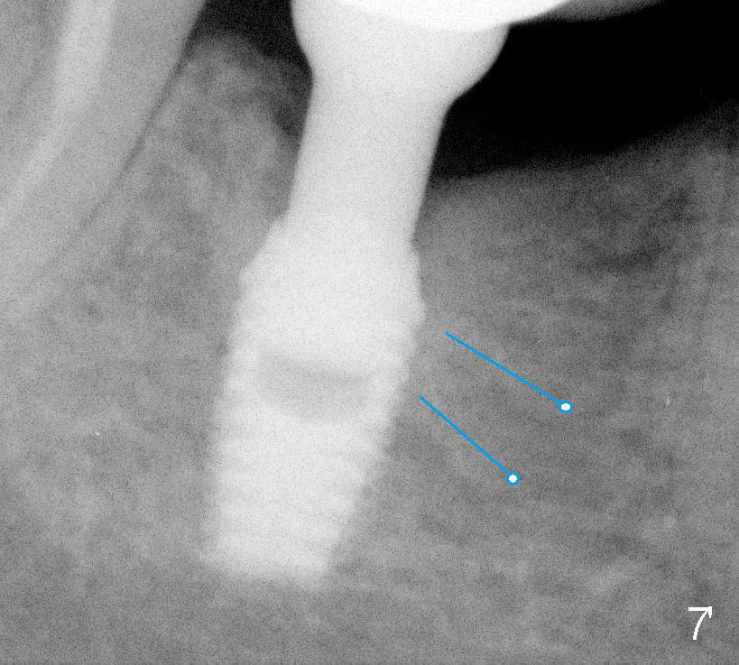
Fig.7 X-ray is taken three years after implant in function. The lower portion of the bone next to the implant has also shown distinct patterns (Fig.7' blue lines). Return to original article
Xin Wei, DDS, PhD, MS 1st edition 04/10/2011, last revision 09/28/2012