 |
 |
|
Fig.1 |
Fig.2 |
Dental Education Lecture: Preparation for Implants
|
|
|
|
Fig.1 |
Fig.2 |
To assure implant success, we need to do some of preparation work: such as comprehensive exam, including X-ray and taking impression. The doctor then pours models. With the X-ray and models, he/she decides how big and how long an implant should be used. With the models, the dentist can make a guide, which helps him/her place an implant during surgery: exactly where to place, what is angulation. Precision is a must.
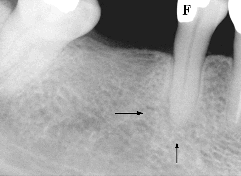
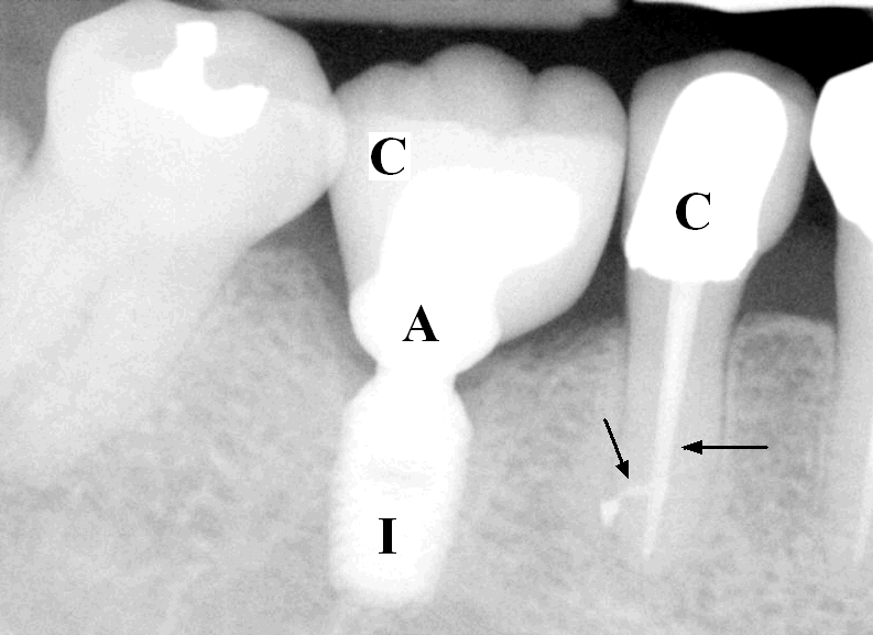
Before elective surgery like implant, we need to have a healthy mouth. Usually you need to have cleaning, filling and root canal done if necessary. We should get rid of any infection nearby implant placement. Fig.1 shows one missing molar tooth. Next to it is a premolar tooth with a large filling (F). The patient complains pain. Careful exam reveals that the nerve is dead. There is root tip infection (two arrows). This type of mild, but persistent infection would adversely affects survival of an implant placed next to it if left untreated. We first finish root canal therapy (arrows in Fig.2), place an implant (I) when pain disappears after root canal, insert an abutment several months later and finally place crowns (C) for both the implant and the natural tooth. It is reported that implant success rate is more than 97% in 7 years. This is due to patients' determination and doctor's good planning and preparation. Do you want to see how an implant-supported tooth looks in the mouth?
Xin Wei, DDS, PhD, MS 1st edition 02/14/2009, last revision 09/28/2012