|
|
|
|
|
|
|
|
Early Hidden Crack of Root
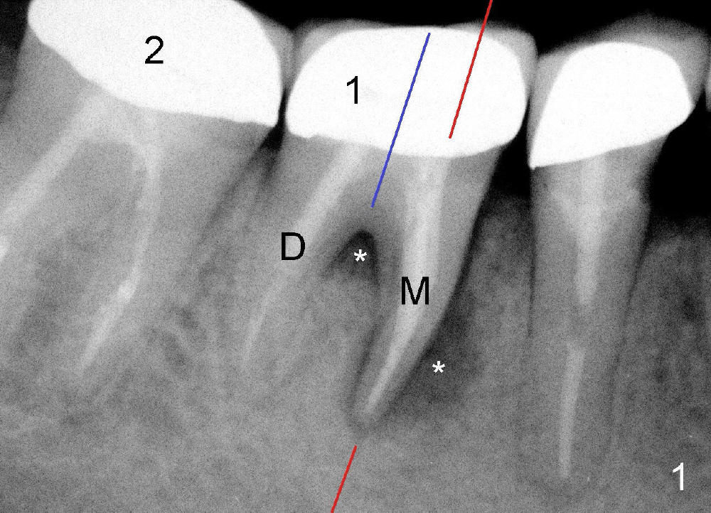
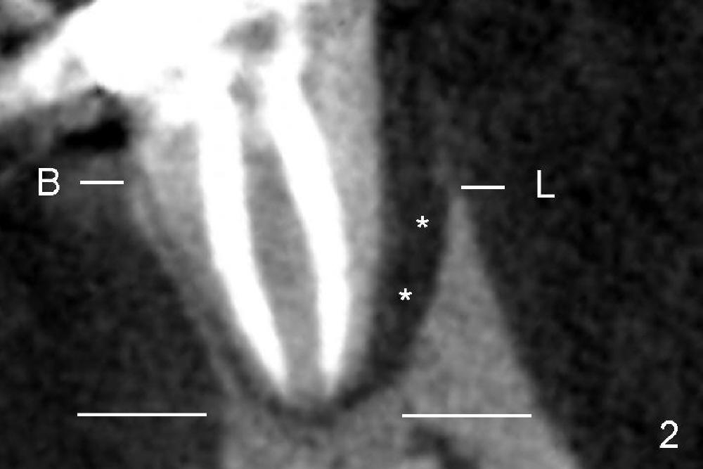
At initial exam, an abscess is found on the tongue side of Mrs. Lei's 1st molar with light tenderness. She is totally unaware of the abscess before exam. X-ray shows that there is bone loss (Fig.1 *) around the mesial root (M, one of the two roots of a lower molar, which is closer to the midline of the face) of the 1st molar (1). Clinical experience tells us that the mesial root has cracked. To confirm the diagnosis, CT scan is taken. Let us make a section through the mesial root along the red line in Fig.1. The CT section shows that there is severe infection (Fig.2 *) on the tongue side of the mesial root (L, lingual (Latin, tongue)). The bone height (between 2 white lines) is lower on the tongue side than that on the cheek side (B: buccal (cheek)). All of these pieces of evidence suggest a possible crack of the mesial root, although there is no visual evidence of the crack from regular X-ray (Fig.1) or CT image (Fig.2). Anyway, the 1st molar should be extracted immediately and replaced by an implant later.
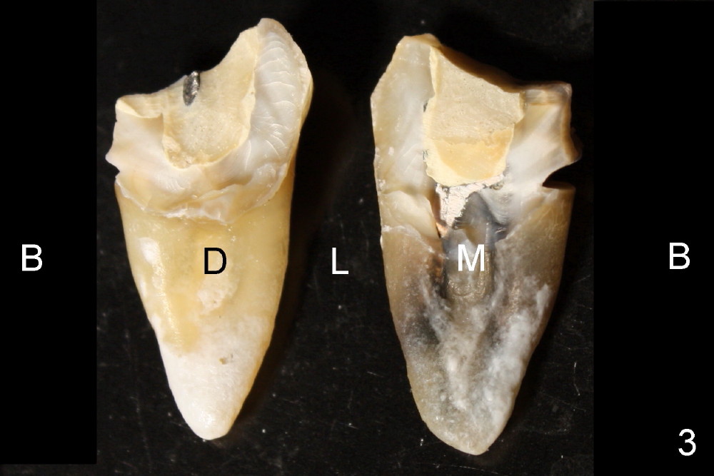
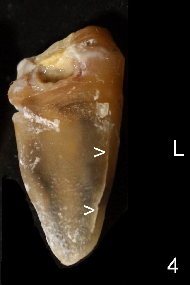
To reduce extraction trauma, the tooth is split along the blue line in Fig.1. When the 2 pieces of the tooth are taken out separately, they are turned 90 degrees: counter-clockwise for the mesial root (M) and clockwise for the distal root (D). These 2 extracted roots are shown in Fig.3. No crack can be found on the mesial root, although it is dark and pretty nasty as compared to the white shiny distal root. When the mesial root is turned for 180 degree, a crack is finally visible (Fig.4 >). The crack is so fine that it is not detectable in CT scan or easily to be missed by a casual look.
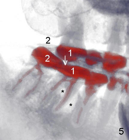
Why does the root of the lower 1st molar crack? The upper 2nd molar on the affected side is missing (Fig.5: 3-D CT image). Almost of all chewing force is transmitted to (arrow) the lower 1st molar with root canal. The tooth that needs root canal is usually in bad shape. After root canal, it becomes even weaker. To strengthen Mrs. Lei's chewing system, she needs two implants to replace the two missing teeth, one on the top and the other on the bottom.
Xin Wei, DDS, PhD, MS 1st edition 08/31/2012, last revision 09/02/2012