
|
|
|
||
|
|
|
||
|
|
|
||
|
|
|
Dental Education Lecture: Implant and Nerve
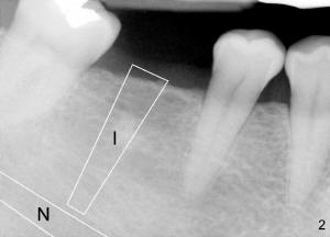
When we plan to fix a missing bottom molar (Fig.1: *) with an implant (Fig.2: I), we need to know where the nerve is (N). We want to have a long implant without touching the nerve. However, regular X-ray does not show the nerve clearly (compare Fig.1,2). There is a lot of guess work. Damaging the nerve during implant surgery will cause severe complications. Our lower lip on affected side remains numb after anesthesia wears off. Therefore, we should eliminate guess work by doing the following.
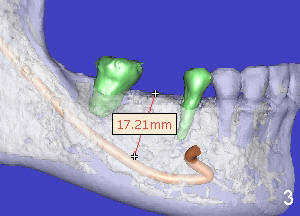
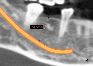
Before implant surgery, it is important to have oral CT (computerized tomography) taken. CT provides 3-dimensional image of our teeth and nerve (Fig.3: brown). Everything is crystal clear. Bone height is measured to determine what the length for the implant is. The measurement can be also done in CT cross section (2-dimension) (Fig.4), similar to regular X-ray shown in Fig.1,2.
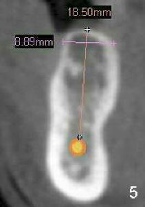
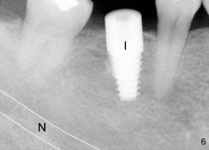
CT transverse section of the lower jaw shows the width of the jaw (Fig.5: 8.89 mm), which decides the diameter of the implant (6 mm in Fig.6). On the safe side, a wider, but shorter implant (6 mm (in diameter) x 14 mm (in length) is placed for this case (Fig.6: I, as compared to Fig.2). There is plenty of space between the implant and nerve. The patient is doing very well during and after surgery. There is no sign of nerve injury.
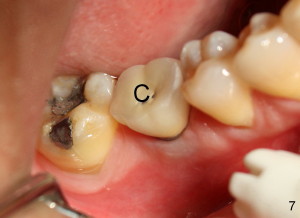
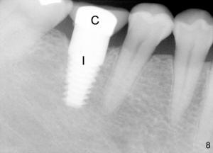
Six months later, she has a new tooth (Fig.7: C, crown). The gums underneath the crown (i.e., around the implant) is healthy. She returns to our office for cleaning six months after getting the new molar tooth. She is pleased to tell us that the implant tooth is no difference from her natural teeth (Fig.8).
Xin Wei, DDS, PhD, MS 1st edition 03/21/2011, last revision 03/21/2011