

|
|
|
 |
| Fig.1 | Fig.2 | Fig.3 |
|
|
|
 |
| Fig.4 | Fig.5 | Fig.6 |
 |
<--Fig.7 |
Dental Education Lecture: Expose Incisor with Laser
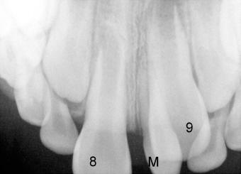
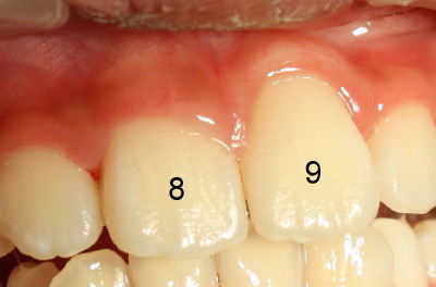
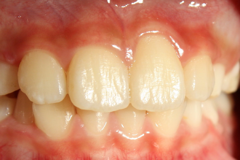
One of Gavin's front teeth (#9 in Fig.1-3 vs. its counterpart: #8) does not come out of gums due to blockage of an extra tooth (so-called mesiodens (M)). So we need to remove this extra tooth, which is also misshaped, like a pyramid. One month after extraction, the impacted tooth (#9 in Fig.4) comes down quite a lot underneath the gums, as compared to Fig.1. Gavin's mom worries about so much that she asks us to bring the impacted tooth out immediately. There is bruise (arrowhead) near the incisal edge of the impacted incisor. With usage of laser, we expose the incisor without any bleeding (Fig.5, as compared to surgery with blade). The other advantage of using laser is that there is much less pain after surgery, because laser has anti-inflammatory effects.
The impacted tooth erupts normally very soon. A year after the last procedure, the central incisors (#9) is at the same height as the neighboring tooth #8 in Fig.6. Gavin's mom is still much pleased with his front teeth (Fig.7, 1 year and 10 months after surgery).
Xin Wei, DDS, PhD, MS 1st edition 06/09/2010, last revision 04/24/2012