|
|
|
| Fig.1 | |
|
|
|
| Fig.2 | Fig.3 |
|
|
|
| Fig.4 | Fig.5 |
|
|
<--Fig.6 |
Dental Education Lecture: Cyst around impacted tooth
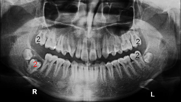
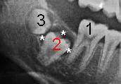
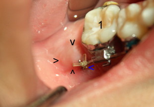
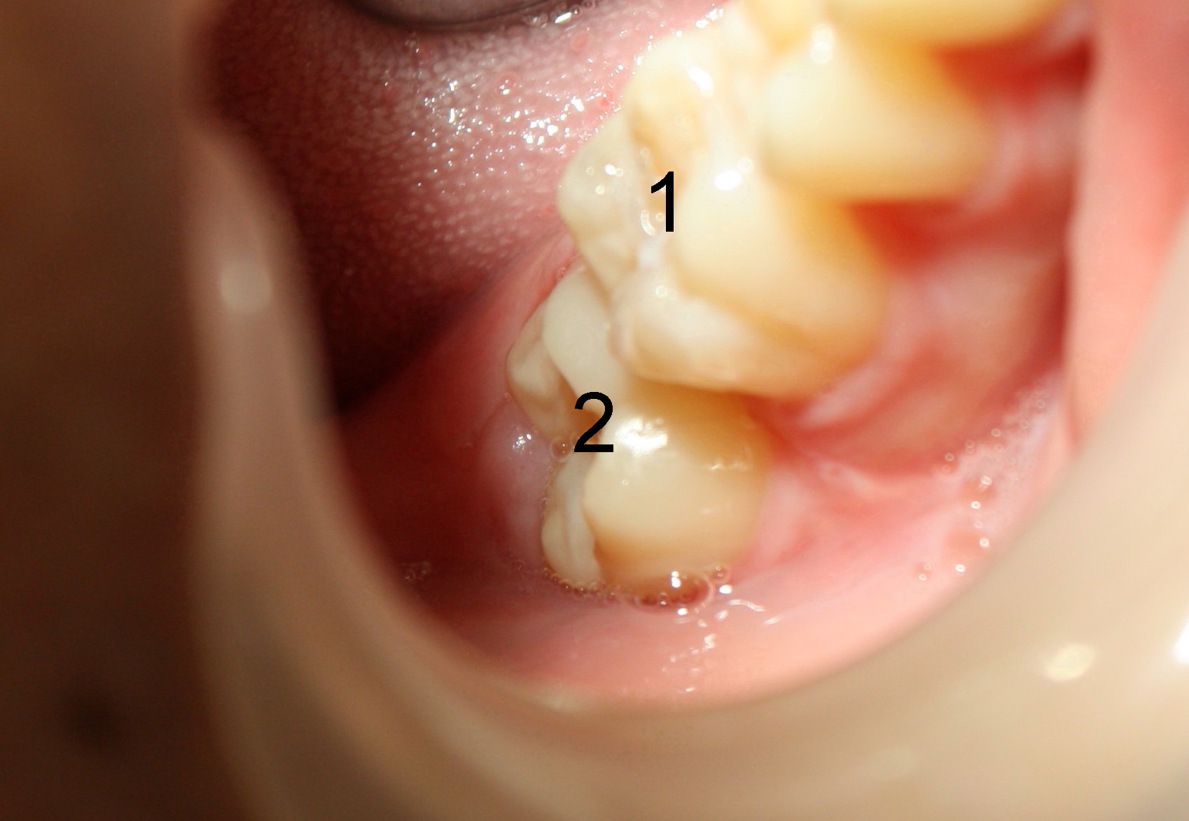
Cyst is a benign tumor that sometimes forms around an impacted tooth. Michael is 13 years old. One of 2nd molars has not erupted (still inside jawbone, Fig.1: red #2), while the other three have (black #2). There is abnormally large space (Fig.2 *) around the impacted molar. By comparison, the space around the third molar tooth bud (3) is normal. The abnormally large space (cyst) prevents the molar (2) from breaking through the bone above. Besides, the third molar (wisdom tooth) may also present an obstruction for the 2nd molar to make appearance into the mouth.
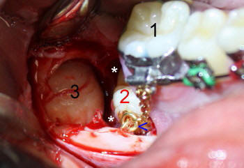
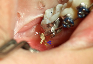
Therefore, we should remove the bone above the impacted 2nd molar, remove the cyst, take out the wisdom tooth and drag the 2nd molar out. Here we go. Fig.3 shows that we have removed the bone and cyst above the 2nd and 3rd molars. Asterisks (*) indicates the abnormally large space, where the cyst was seated. One end of a gold chain (blue arrowhead) is attached to the impacted 2nd molar. The other end of the chain is fixed temporarily to braces of the 1st molar (1). Finally the 3rd molar (3) is extracted. Michael recovers smoothly after surgery. Four weeks after surgery, the wound heals (Fig.4). The orthodontist (Dr. Tracey Salyer) uses a small rubber band to tie the gold chain (blue arrowhead) to the metal wire above (white arrowhead) to facilitate the eruption of the impacted 2nd molar. Another six months later, Michael returns for cleaning. The impacted 2nd molar almost makes its appearance. You can see a bulging area (Fig.5 black arrowheads), just like a flower almost blossoming. Another hint of the tooth coming out is that the gold chain is shorter in Fig.5 than that in Fig.4 (blue arrowhead). The thicker wire above is bent (compare Fig.4,5 white arrowhead), suggesting that it has a lot of tension and tries to pull the impacted tooth upward with the help of elastic (rubber band). One year after surgery, the impacted 2nd molar makes its appearance (Fig.6: 2).
Xin Wei, DDS, PhD, MS 1st edition 01/10/2011, last revision 04/21/2012