|
|
Mucocele Formation and Treatment
Although mucocele is a benign lesion (cyst), it is bothersome. It keeps coming back. It needs surgery. In this lecture, we are going to use a diagram to demonstrate how mucocele forms and why surgery not only remove the cyst, but also its roots.
As we know, our mouth has saliva, water-like mucus. Saliva wets our mouth so that we can talk, eat and swallow. Saliva is produced by salivary glands outside our mouth and underneath oral mucous membrane.
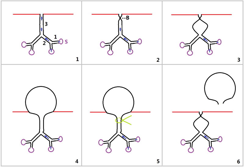
Fig.1 shows that underneath mucous membrane (red line) is a salivary gland. The latter consists of several mucus-producing sacs (S: purple circles, like grapes) and ducts (black tubes). After formation, mucus is emptied (arrow) into the 1st segment of salivary duct (1 in Fig.1). Several (two in the diagram) of the 1st segments of ducts join and empty mucus into the 2nd segments of duct. Then several of 2nd segments of ducts join and empty mucus into the 3rd segment of duct. Finally mucus is dumped into the oral cavity (above the red line).
Every day, we eat and bite several hundred times. Occasionally we may bite our lip. The opening of the salivary duct is blocked (Fig.2 B) so that mucus is accumulated inside the salivary ducts and sacs. A small balloon forms (Fig.3). With time, the balloon becomes big and bulges into oral cavity (Fig.4).
The cyst is painless. We love to bite on it or use a string (Fig.5 green line) to pinch it. After water-like mucus leaks out, the cyst disappears (Fig.6). But the opening may remains blocked. Mucus builds up again inside. The small balloon makes comeback and eventually bulges again into oral cavity (as shown in Fig.4).
To eradicate the cyst, the whole salivary gland including several sacs needs to be removed as well (Fig.4).
Xin Wei, DDS, PhD, MS 1st edition 04/10/2012, last revision 04/10/2012