
 |
 |
 |
|
 |
 |
 |
 |
Delayed Pain after RCT Retreatment
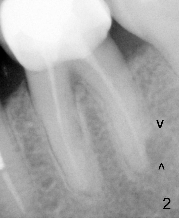
Mrs. Yuan presented to my clinic in March 6, 2007 with chief complaint "dull pain in lower left after root canal". It appears that there is resorption involving the distal root of #19 with periapical radiolucency (PARL, Fig.1,2 black arrowheads). There is open margin involving the crown (white arrowhead in Fig.1). Although there is moderate bone loss (Fig.1), oral hygiene was maintained very well.
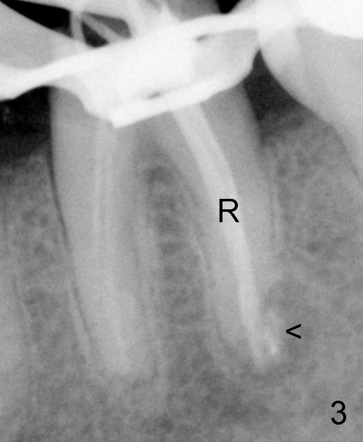
Two months later, after removal of the existing crown, RCT retreatment was initiated in the distal root (Fig.3: R (lateral condensation: master cone 40/.06 with overfilled paste: arrowhead). The access to the mesial canals is extremely limited; gutta percha cannot be removed from the mesial canals with Chloroform. Since the periapical lesion with mesial canals is not a major issue, the mesial canals are left without retreatment.
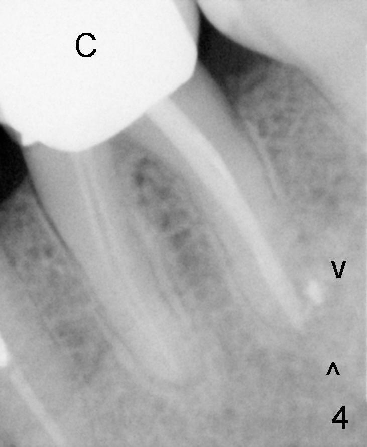
Six months after RCT retreatment, follow-up PA shows reduction in PARL distally (Fig.4 arrowheads) with improvement of symptoms as well. Also notice the new crown (C) without open margin.
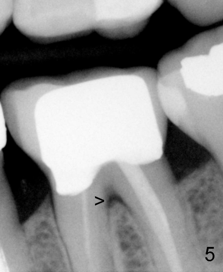
One year later (January 19, 2009), bitewing shows the earliest sign of furca radiolucency (Fig.5 arrowhead). The crown margin remains intact.
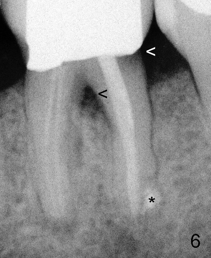
On January 18, 2011, Mrs. Yuan returns to my office with discomfort in lower left. Clinical exam shows 2nd caries in the distolingual line angle, which is confirmed by periapical film (Fig.6 white arrowhead). Furca lesion appears to enlarge (black arrowhead, as compared to Fig.5). Mrs. Yuan's oral hygiene deteriorates due to her general health.
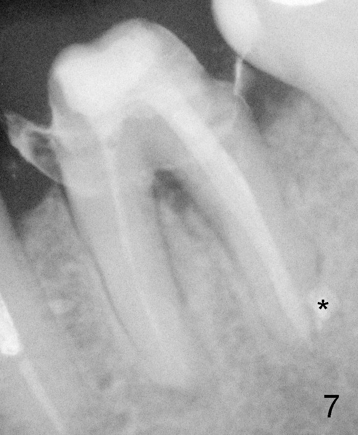
After removal of the existing crown and carious lesion and placement of a temporary crown (Fig.7), probing pain is more or less concentrated on lingual furcation. Distal PARL disappears around overfilled paste (* in Fig.6,7).
Please evaluate the tooth #19 for diagnosis and further treatment. Thank you for consideration.
Xin Wei, DDS, PhD, MS 1st edition 07/20/2011, last revision 03/23/2018