
 |
 |
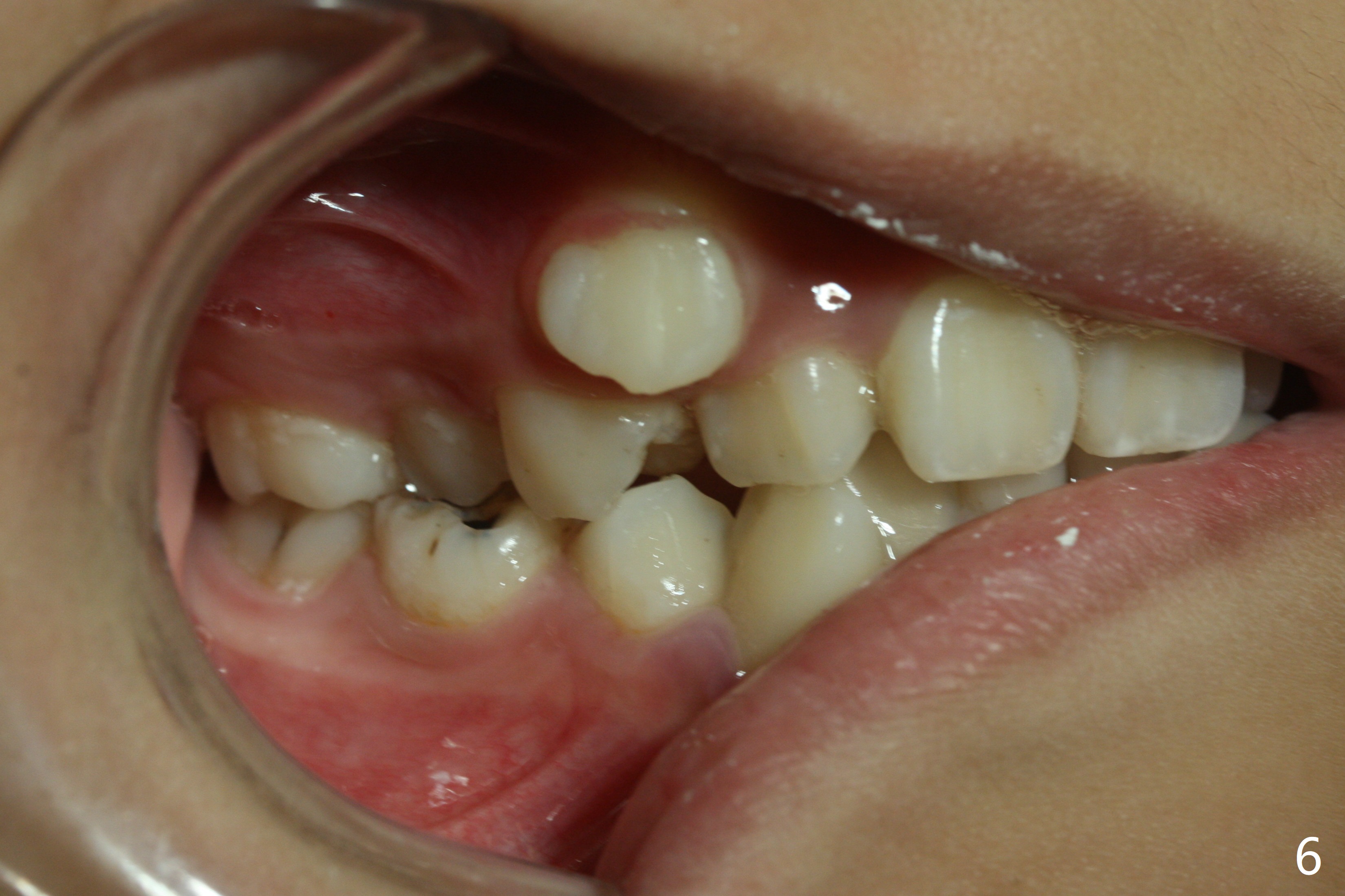
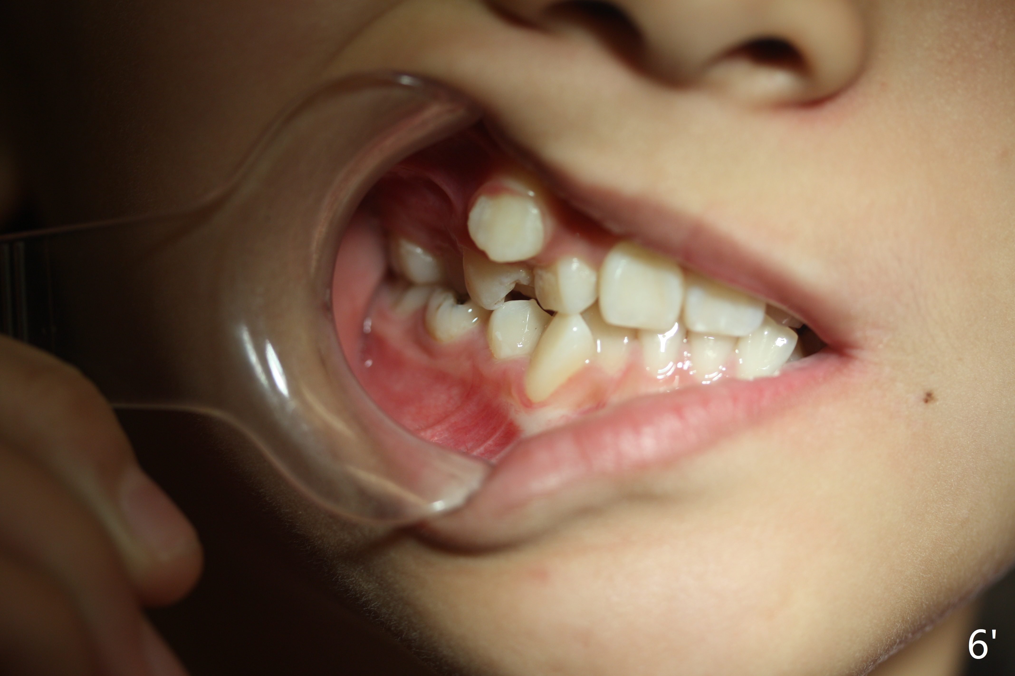
The lateral view of occlusion is to show the relationship between the upper and lower molars (Fig.6): Class I, II or III occlusion. The axis of vision should be perpendicular to the plane of interest. Otherwise Fig.6' is no difference from the previous one. Ask the patient or somebody else to retract the lip. You place the camera in the right way.
Xin Wei, DDS, PhD, MS 1st edition 07/29/2018, last revision 08/08/2018