|
|
|
|
|
|
|
|
|
Implant is not Nude in the Sinus
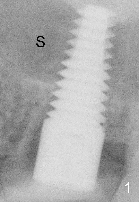
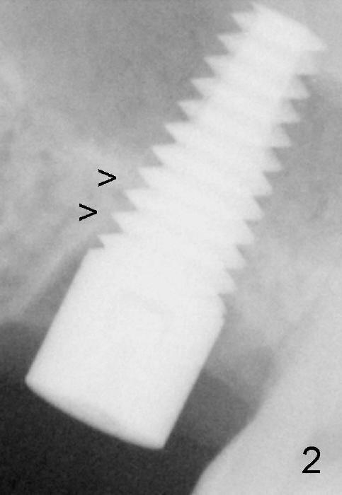
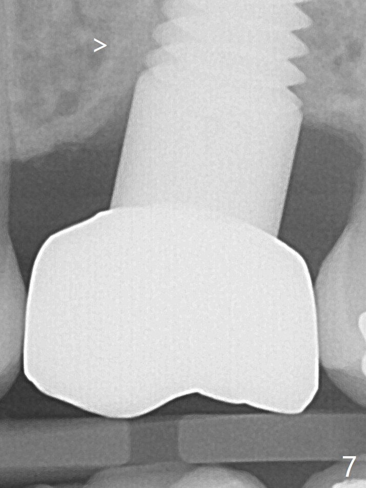
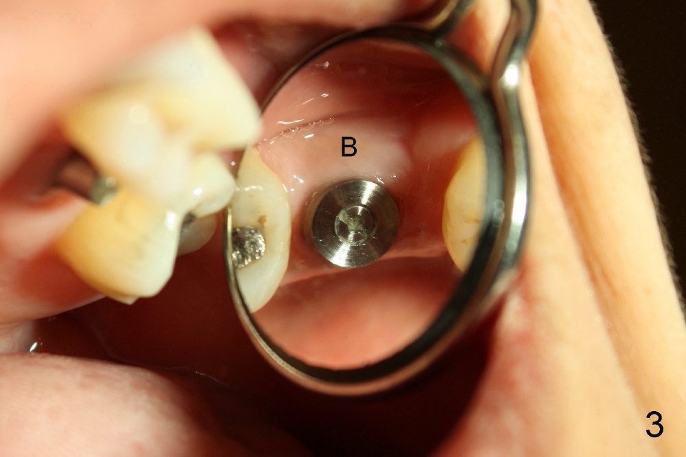
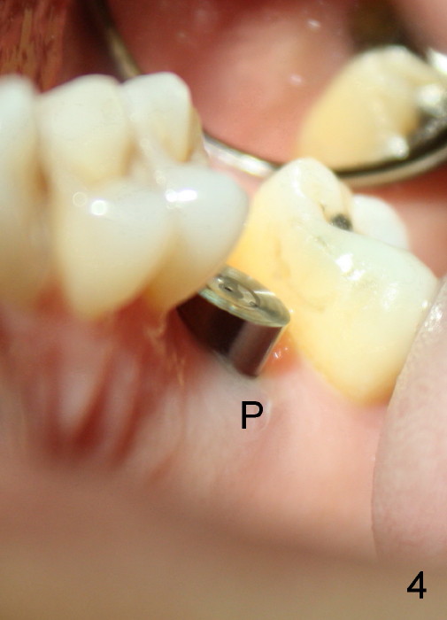
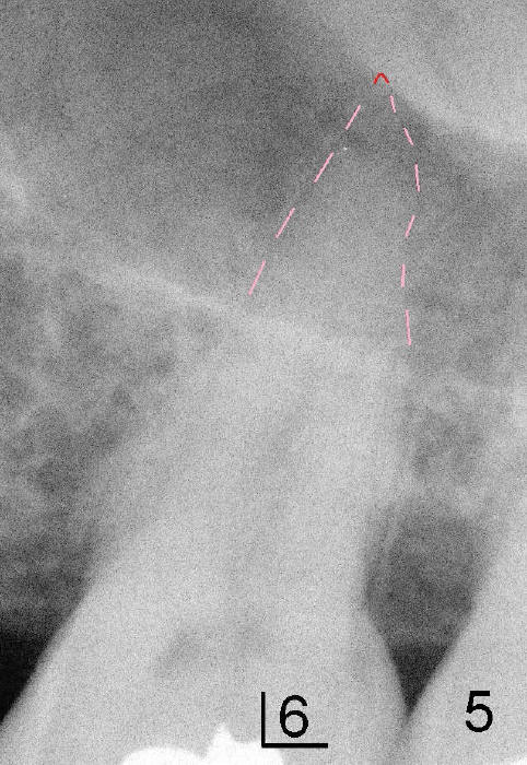
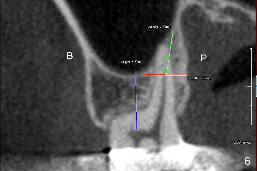
Prior to placement of 6x17 mm soft tissue level implant (Fig.1), the same size of tap is used. When the latter is removed, there is a small defect in the buccal wall of the osteotomy (mainly in the palatal socket) with the intact sinus membrane. There is no intraop or postop nasal hemorrhage. The patient returns for restoration 7 months postop; it appears that there is osteo-integration (Fig.2 with the mesial gap getting smaller (>)). The gingiva is healthy buccal (Fig.3: B) and palatal (Fig.4: P). The mesial gap is closed with formation of dense bone 4 years post cementation (Fig.7). Fig.5 is the preop PA of the patient, which shows that the palatal root (pink dashed line) is above the sinus floor and surrounded by the lamina dura. Fig.6 is a coronal section of the 2nd molar of another patient, which shows that the palatal root is 5-6 mm above the sinus floor. Upper Molar Immediate Implant Follow-Up Xin Wei, DDS, PhD, MS 1st edition 10/13/2013, last revision 05/18/2018