|
|
|
|
|
|
|
|
|
|
|
|
|
|
|
3 Steps of Grafting
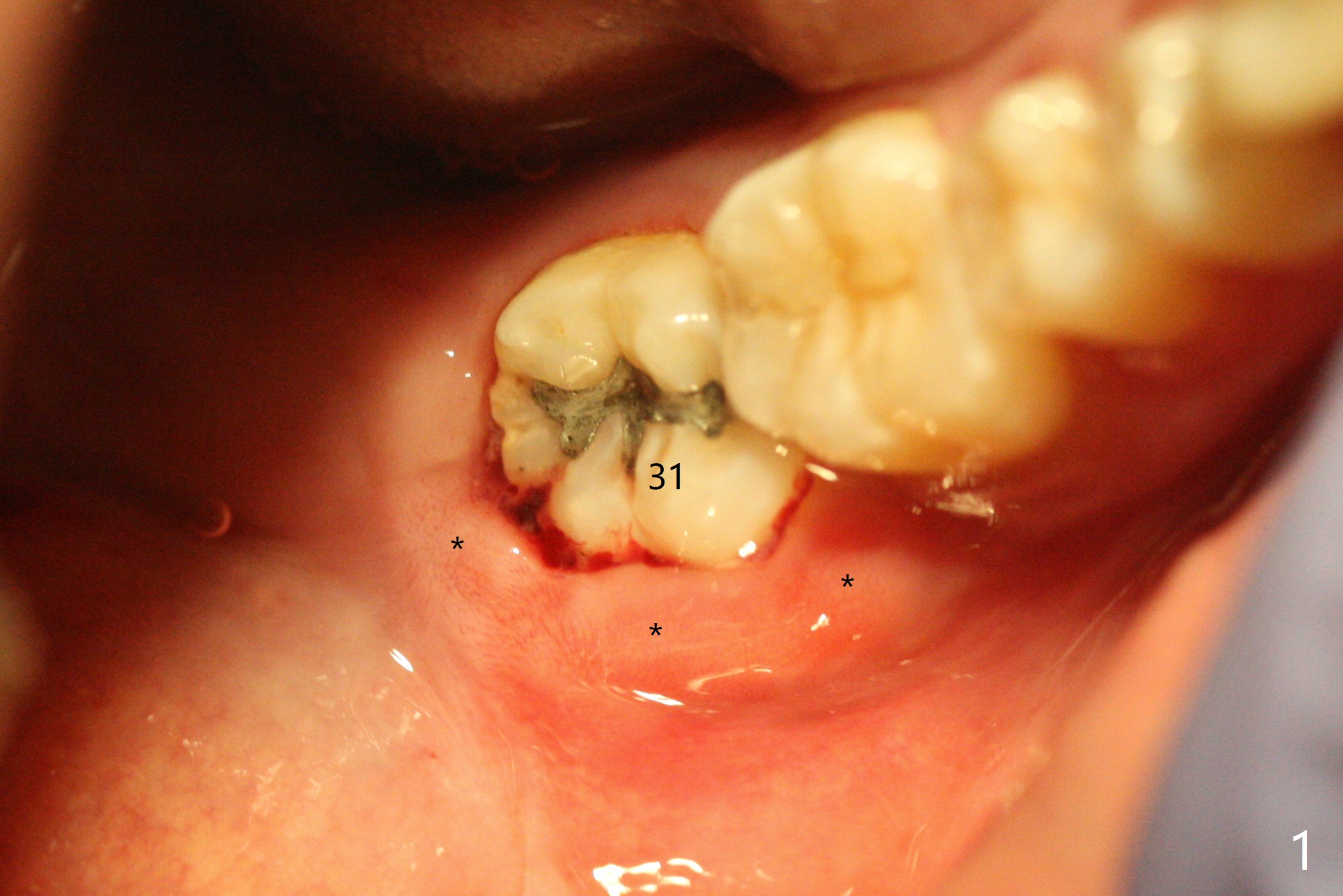
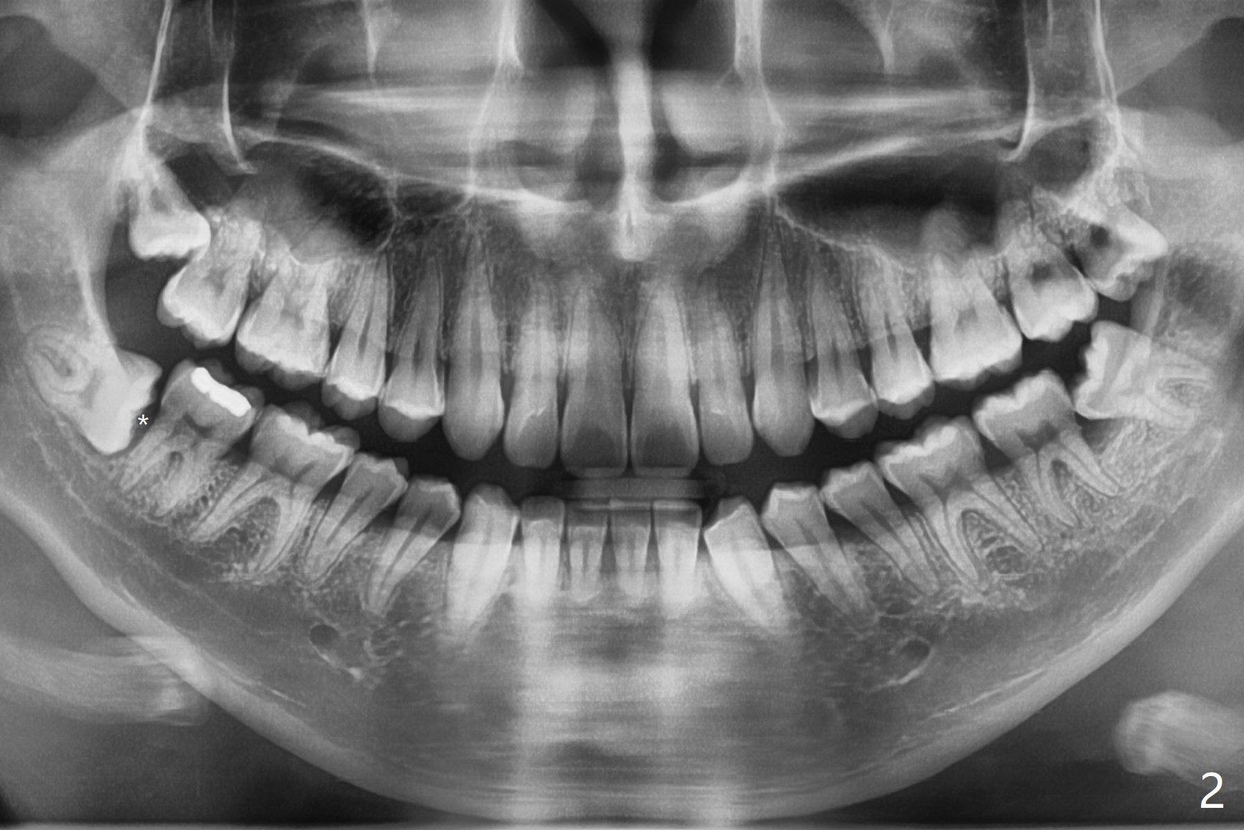
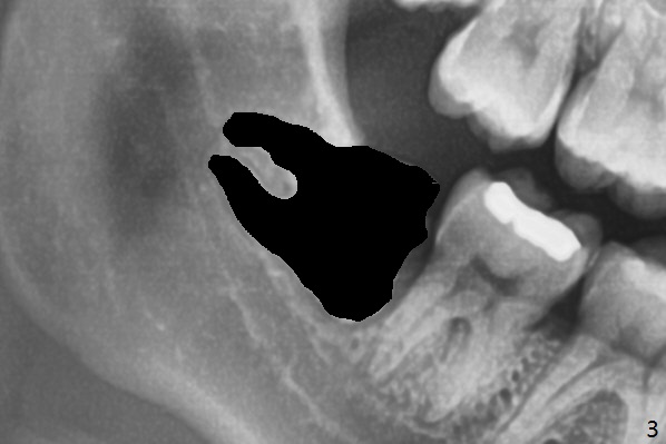
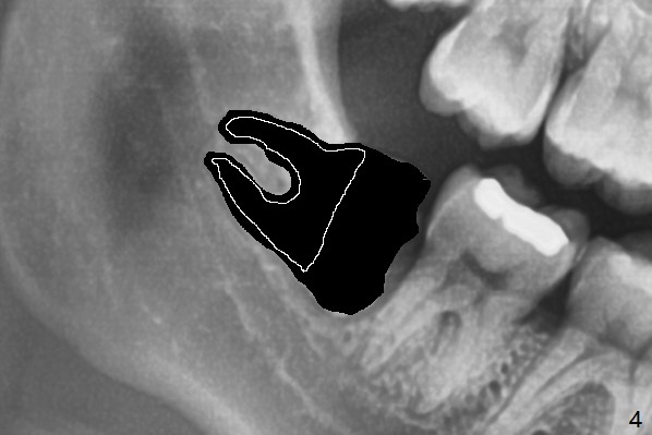
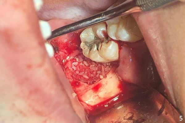
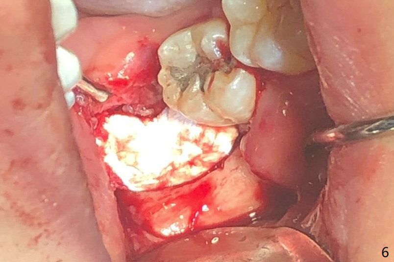
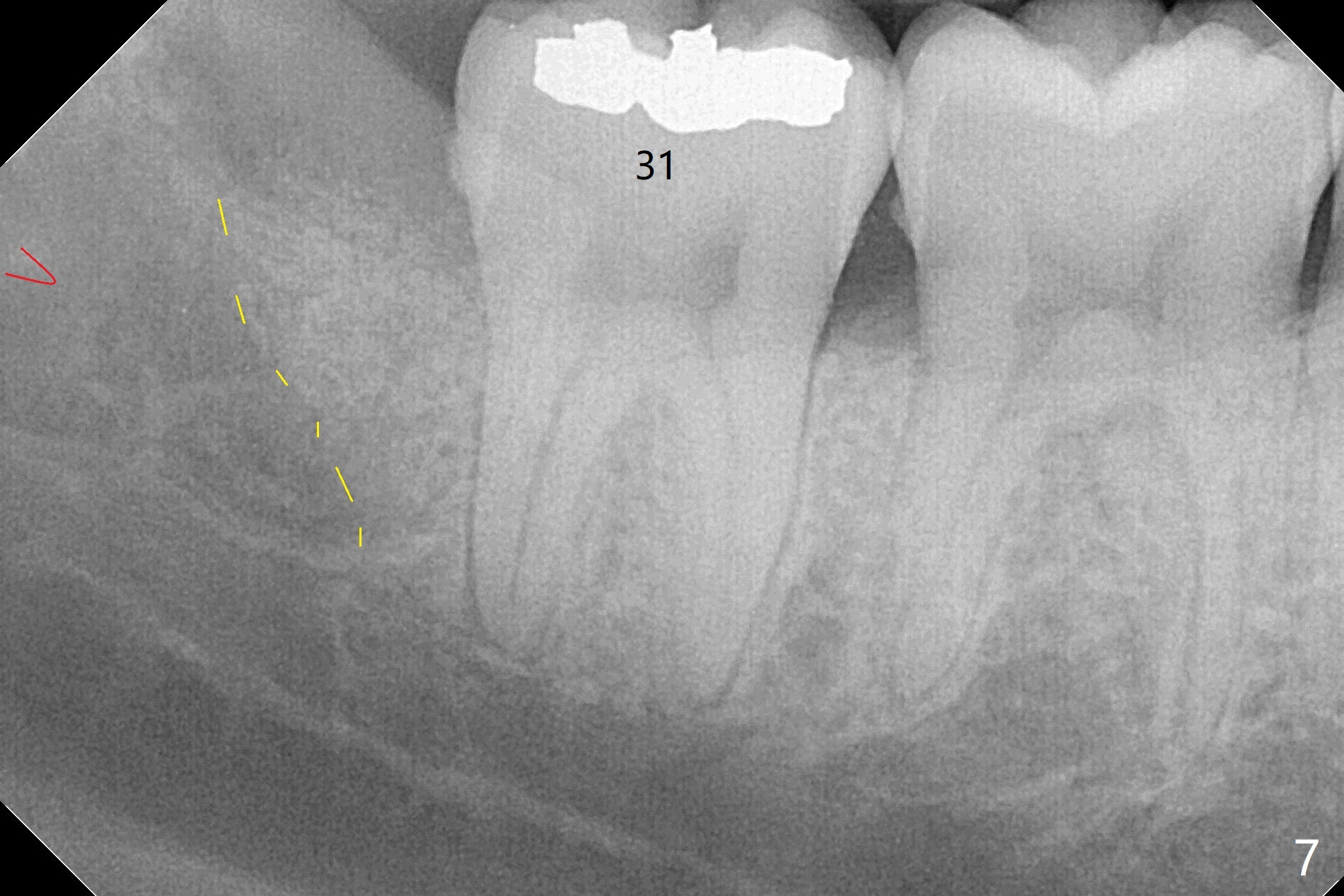
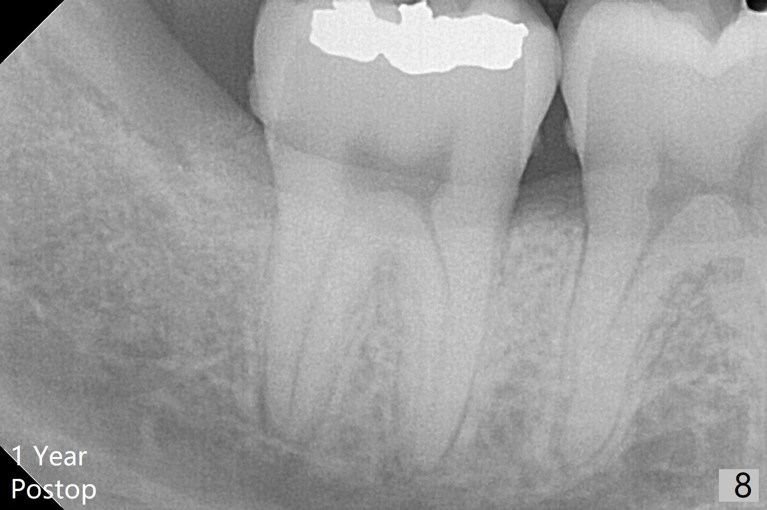
A 22-year-old man with poor oral hygiene requests extraction of the lower right 3rd molar. The buccal gingiva of the 2nd molar is erythematous and edematous (Fig.1 *). There is tenderness between #31 and 32 with severe bone loss (Fig.2 *). After extraction and debridement (Fig.3), Osteogen plug is inserted into radicular portion of the socket (Fig.4 white outline), while allograft is placed coronally (Fig.5, 7), covered by Collagen plug (Fig.6) before suturing. The bone fills the whole socket 1 year postop (Fig.8); there is no root surface exposure at #31. The bone fills the whole socket of #32 one year postop (Fig.8); there is no root surface exposure at #31.
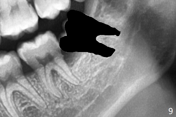
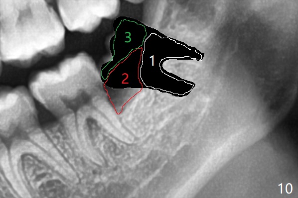
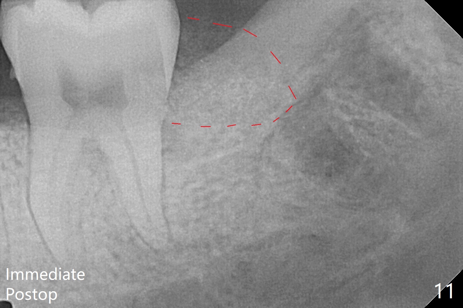
When the tooth #17 is extracted (Fig.9; 23 years old), collagen plug is inserted into the root portion of the sockets (Fig.10: 1 (white outline)), Vanilla graft is placed against the distal surface of #18 (2 (red outline)) and Osteogen plug (3 (green outline)) is placed coronally (3 steps). There is no bony defect associated with the distal surface of the tooth #18 immediate postop (Fig.11 (red dashed line: bone graft)). The wound heals in 2 weeks.
Return to
Plug
智齿拔除
Xin Wei, DDS, PhD, MS 1st edition
08/14/2019, last revision
07/17/2021