|
|
|
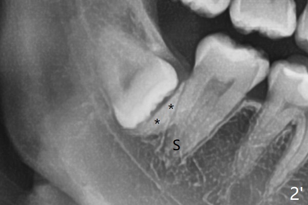
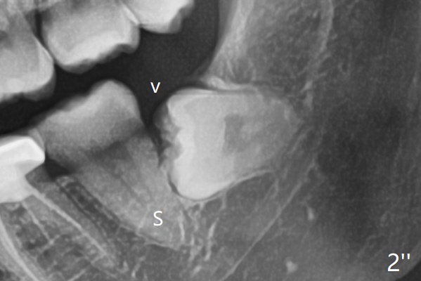
The lower 2nd molars appears to have a single fused root (Fig.2',2'' S); the radiopaque image mesial to the tooth #32 must be the dense bone (Fig.2' *). In contrast the cortex coronal to the tooth #17 is lacking (Fig.2'' v), as compared to that in Fig.2'. It seems that the bone loss at #17 is associated with mild pericoronitis. The tooth #17 should be extracted with placement of Bond Apatite to repair #18 distal defect.
Xin Wei, DDS, PhD, MS 1st edition 06/20/2020, last revision 05/28/2021