


|
|
|
|
|
|
|
The autogenous bone (harvested from reamers), mixed with Bicon Synthograft, is placed in the buccal and mesial portions of the socket prior to placement of a 4.5x20 mm implant (Fig.3 I; 40 Ncm). A: 3.5x5 mm abutment. *: socket gaps.
The mesial and distal socket gaps appear to have been filled by new bone (Fig.8 arrowheads) 5 months postop.
Bone density around the 1st three threads of the implant is particularly high 1 year 3 months post cementation (Fig.10 arrowheads).
By nearly 2 years post cementation, the bone density around the coronal portion of the implant is so high that apparently cortical bone forms (Fig.11 *, lamina dura).
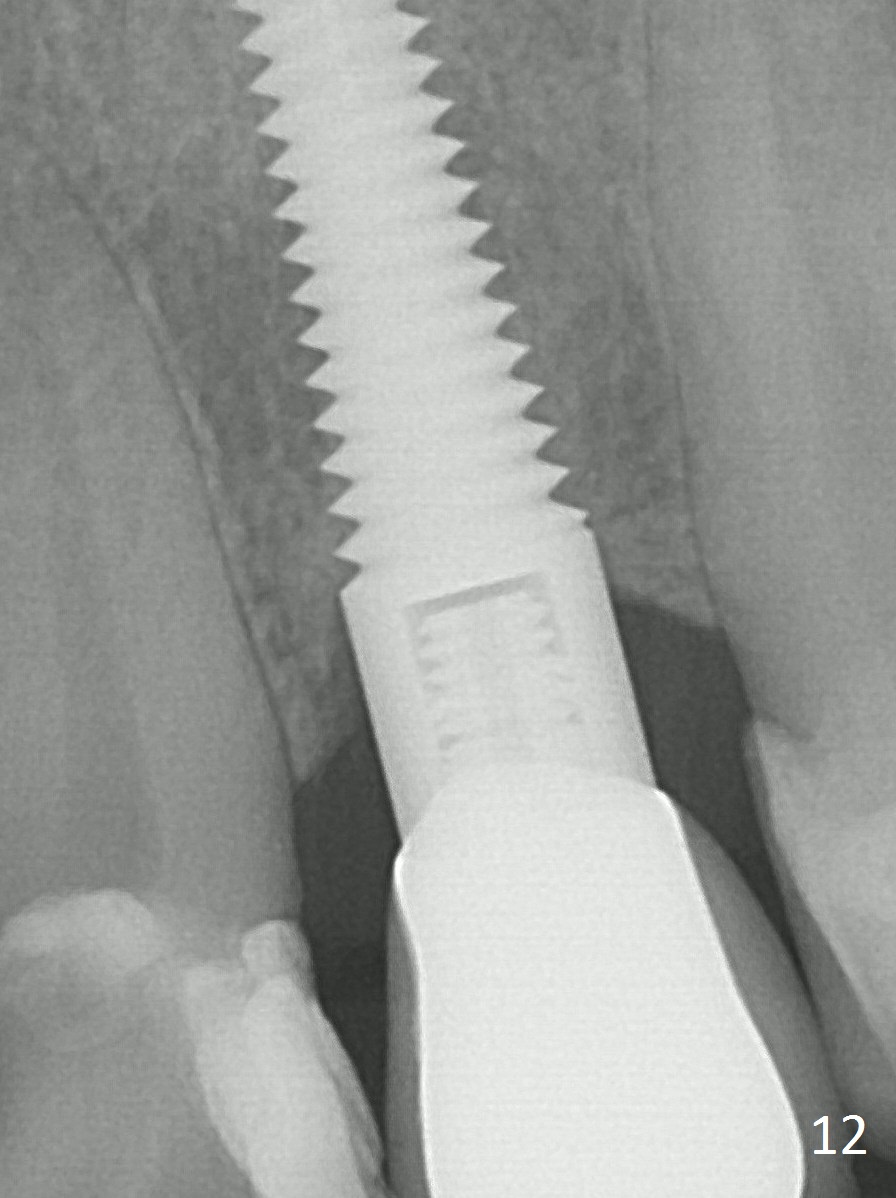
Fig.12: 3 years post cementation.
Possible Osteoporosis for Male
Xin Wei, DDS, PhD, MS 1st edition 08/01/2015, last revision 01/19/2018