|
|
|
PRF and Bone Graft Repair Buccal Plate
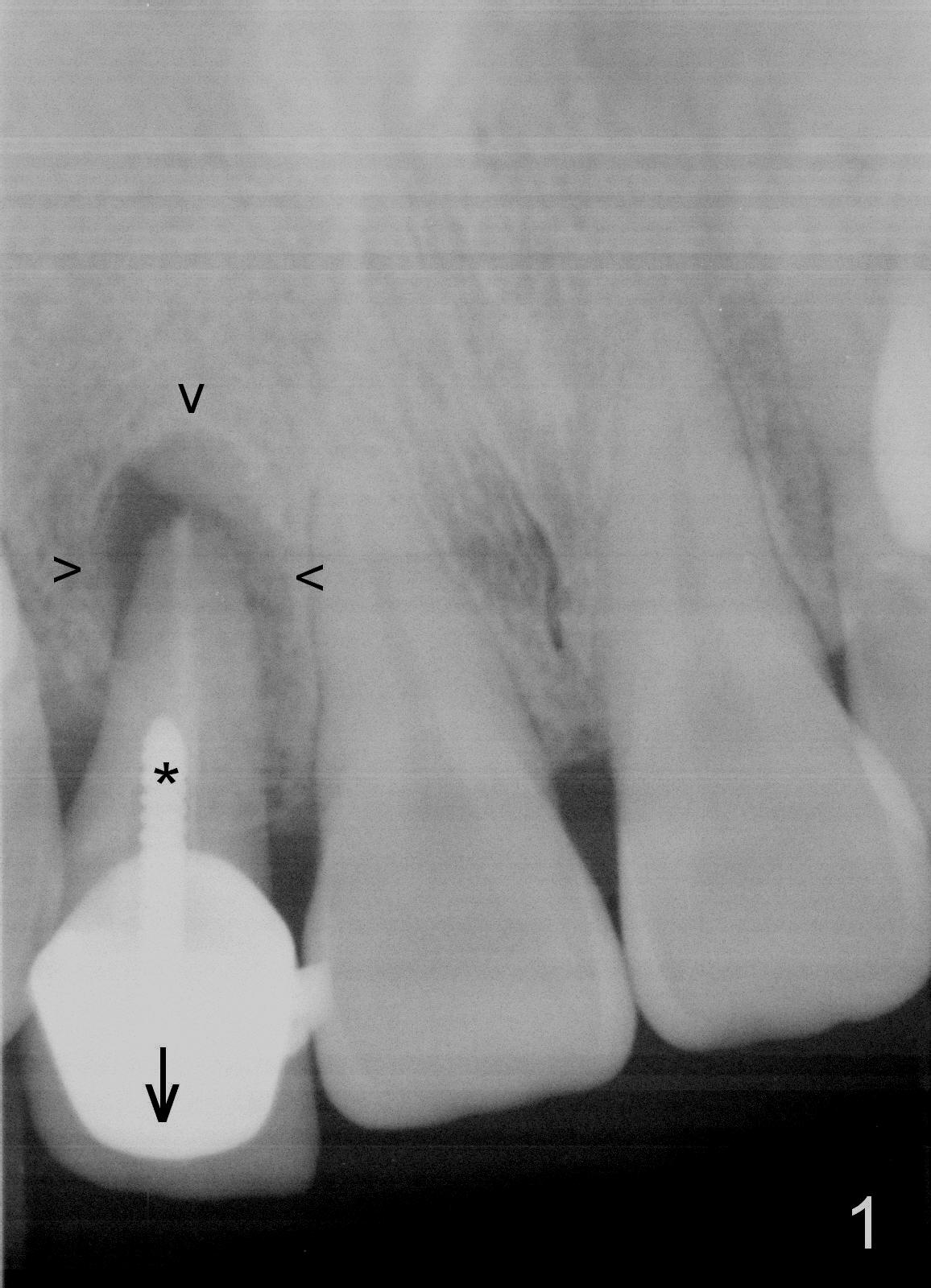
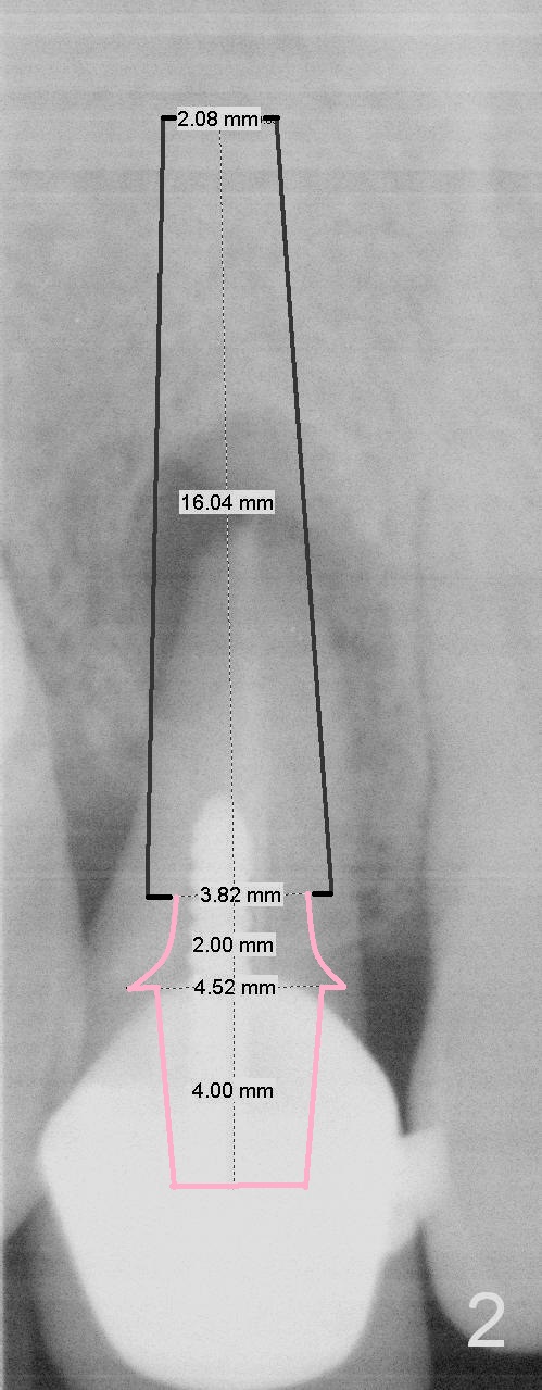
A 42-year-old woman notes that the tooth #7 is extruded (Fig.1 arrow (take preop photo)). It is probably due to buccal plate defect (take intraop photo with curette inside the socket pushing out after extraction). The evidence includes a post (*) and large periapical radiolucency (arrowheads). After Clindamycin socket treatment and implant placement with healing screw (Fig.2), PRF membrane is placed inside the socket against the buccal plate and bone graft is placed between the PRF and the implant. After placement of a cemented abutment (Fig.2 pink), bone graft is placed in the remaining space (2-step grafting), followed by collagen plug or dressing. The existing crown can be modified as an immediate provisional.
Return to
Upper Incisor Immediate Implant
Xin Wei, DDS, PhD, MS 1st edition 04/16/2016, last revision 09/19/2018