.jpg)
%20before%20bone%20graft.jpg)

|
|
|
|
|
|
|
|
|
|
|
|
|
|
|
|||
Exostosis Maintains Buccal Contour
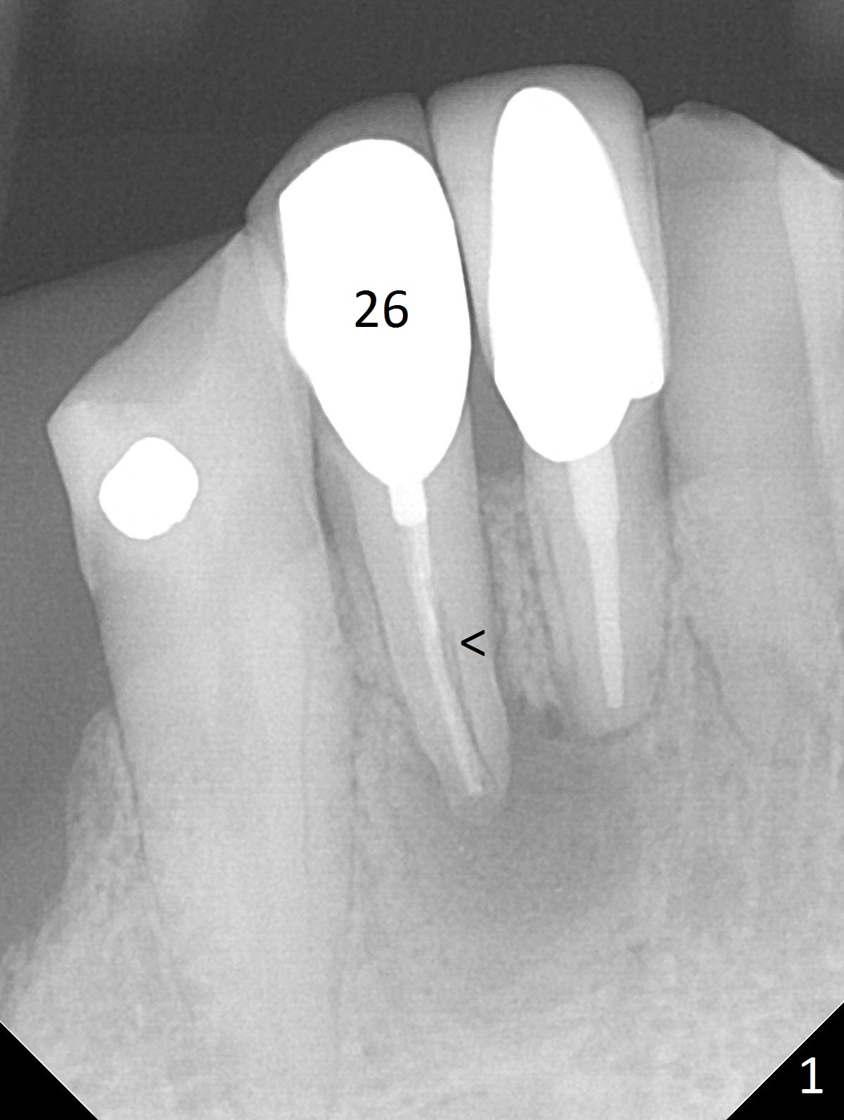
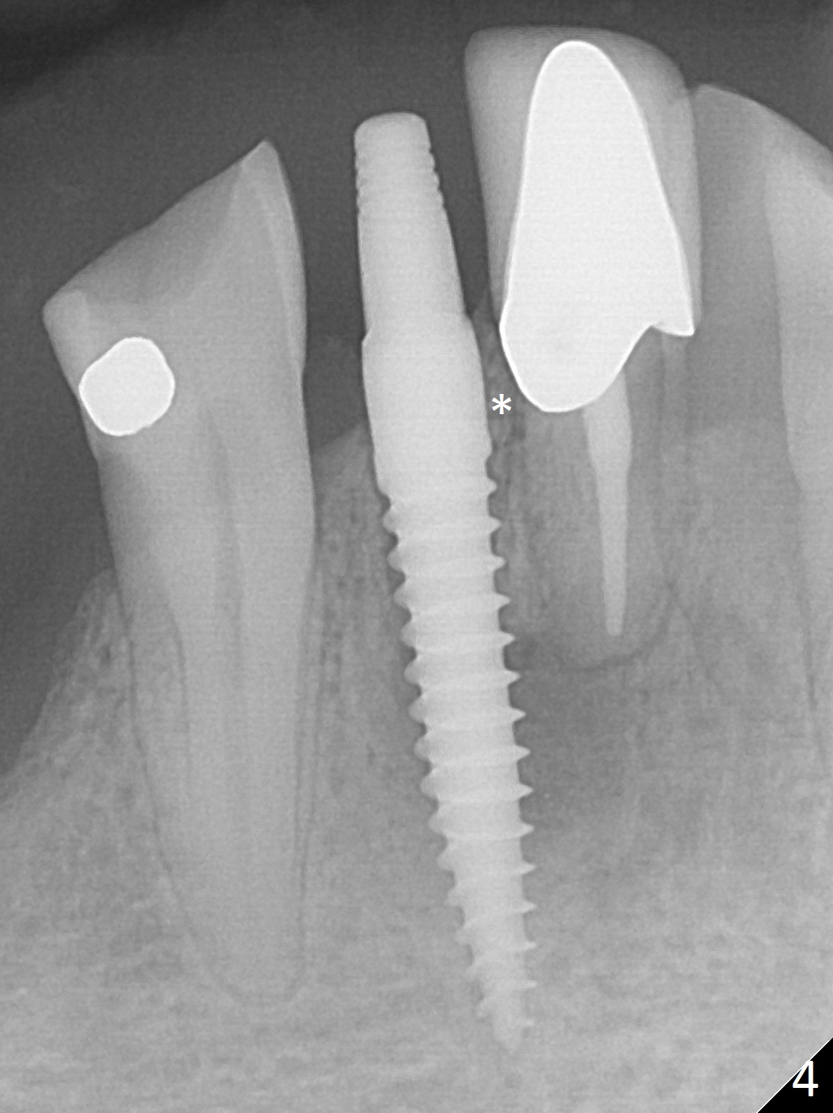
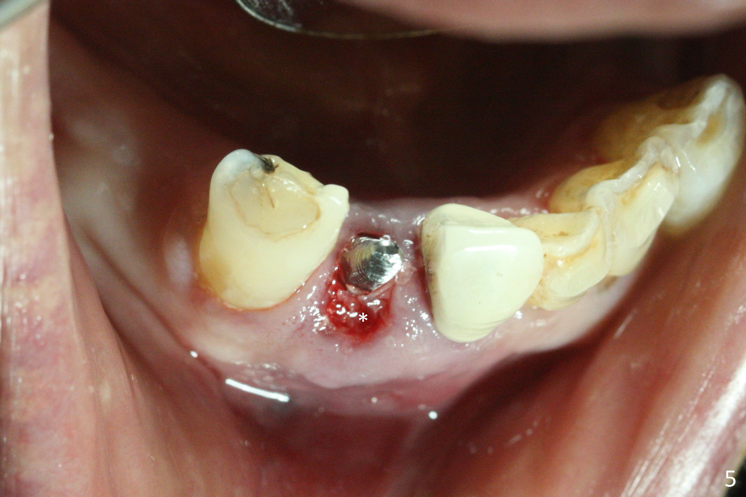
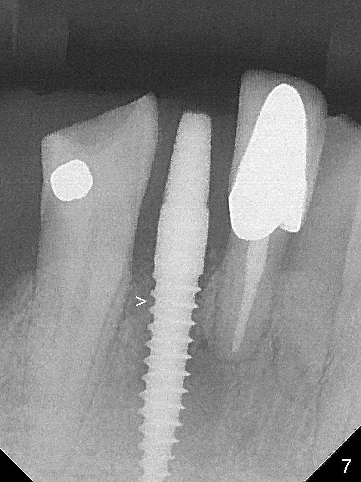
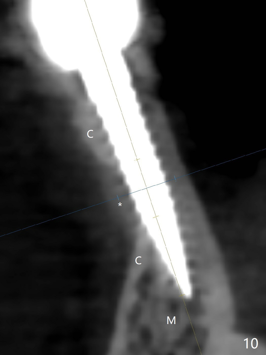
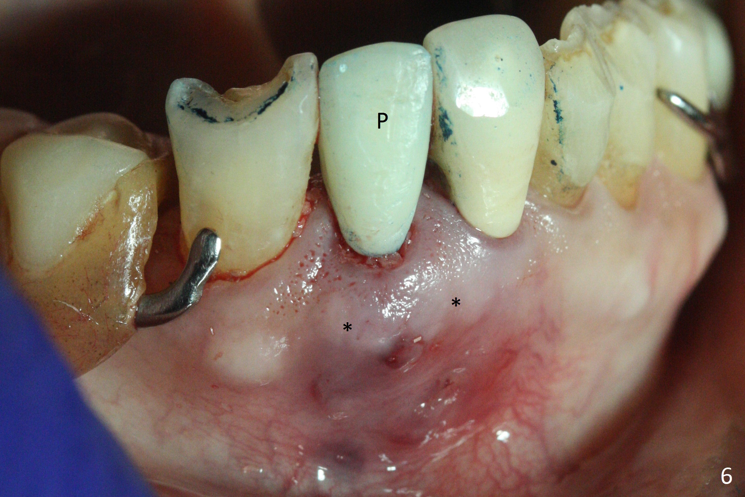
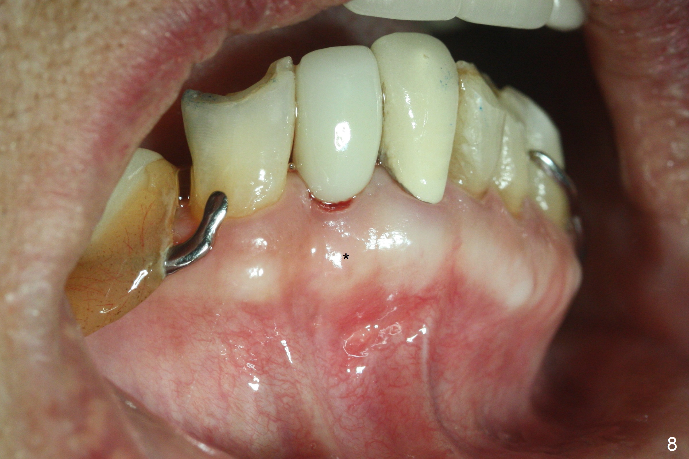
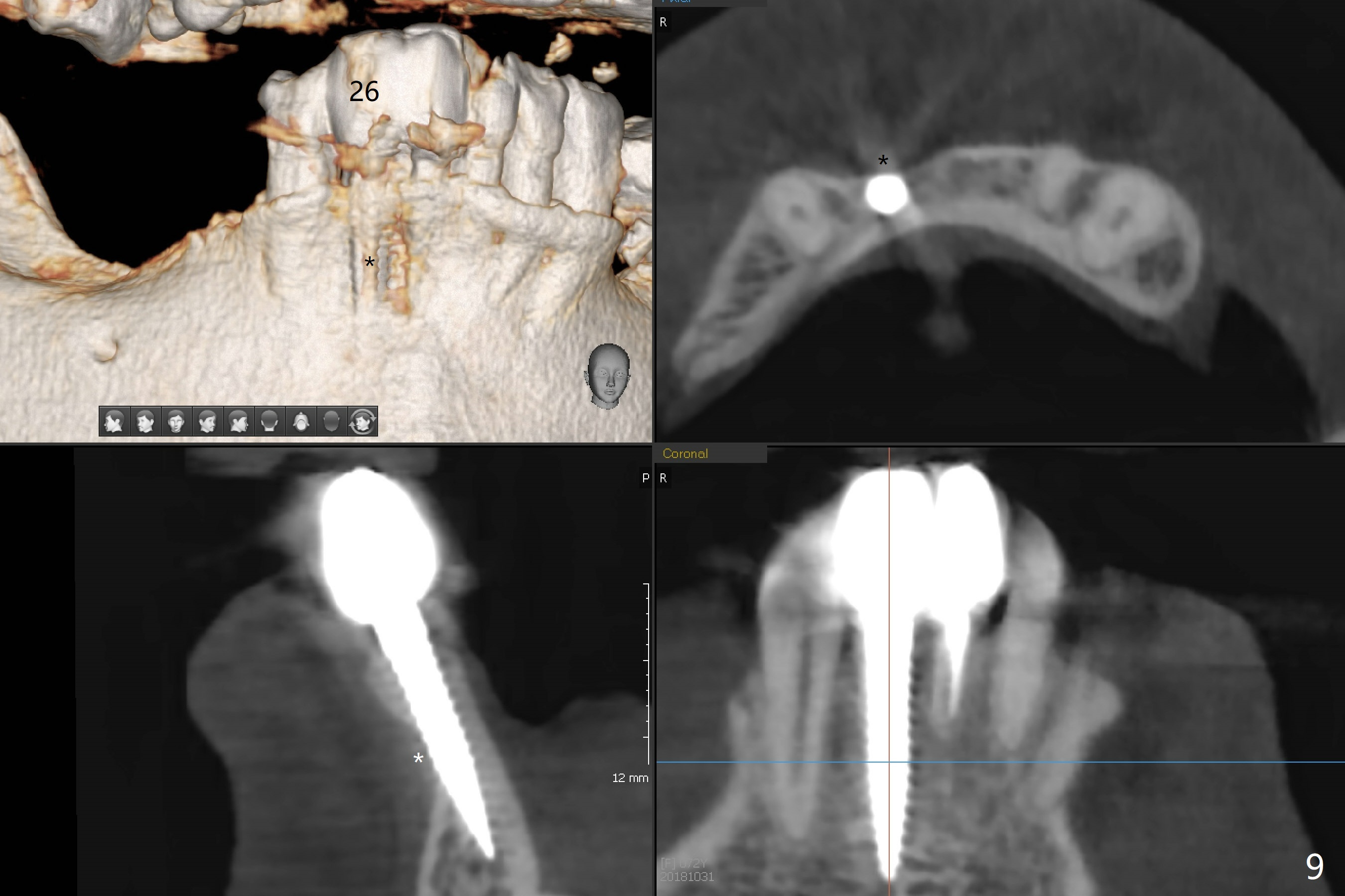
Preoperative X-ray reveals not only large periapical radiolucency, but also vertical root fracture at #26 (Fig.1 <). When the tooth is extracted, the buccal plate is absent except a narrow strip of exostosis (Fig.6 *). A 3x16(2) mm 1-piece implant (Fig.3) is placed lingually (Fig.5) as planned (Fig.2). The socket contour (i.e., buccal gap), most likely maintained by the exostosis, is filled with bone graft (Fig.4,5 *). An immediate provisional holds the graft in place (Fig.6). The distal bone appears to have grown into the space between implant threads in 2.5 months postop (Fig.7 >, as compared to Fig.3). There is no bone loss at the level of exostosis when a permanent crown is cemented 3.5 months postop (Fig.8 *). It appears that the exostosis and bone graft prevents buccal plate collapse. There seems to be new bone formation in the buccal plate (previous PARL and fistula) 1 year 1.5 months post cementation (Fig.9 (CBCT) *). In fact the average density of the new plate (Fig.10 *: 1900 HU) is between the cortical (C: 2600 HU) and medulla (M: 1000 HU) bone. The buccal plate maintains its concavity (Fig.11).
Return to
Lower Incisor
Immediate Implant, IBS,
3, 11-15
Xin Wei, DDS, PhD, MS 1st edition 04/25/2017, last revision 10/31/2018