
|
|
|
|
|
|
|
|
|
|
|
|
|
|
|
|
|
|
Dual Surgical System
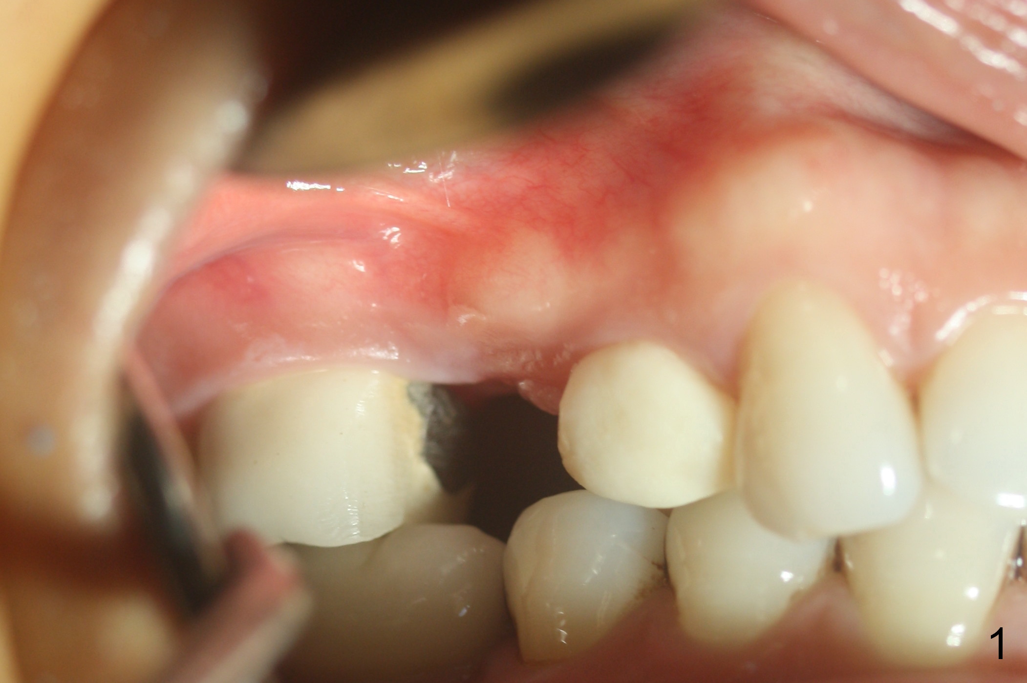
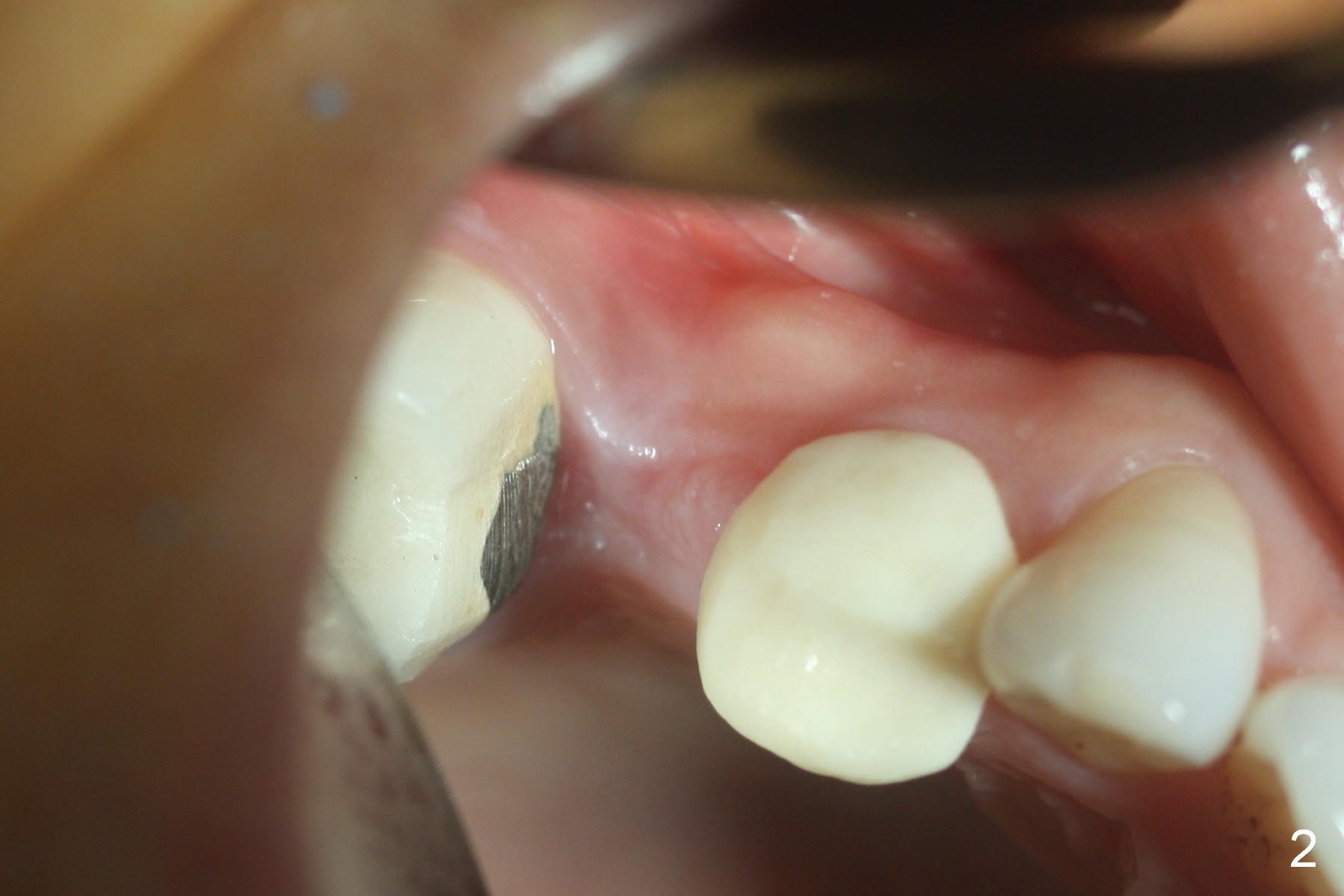
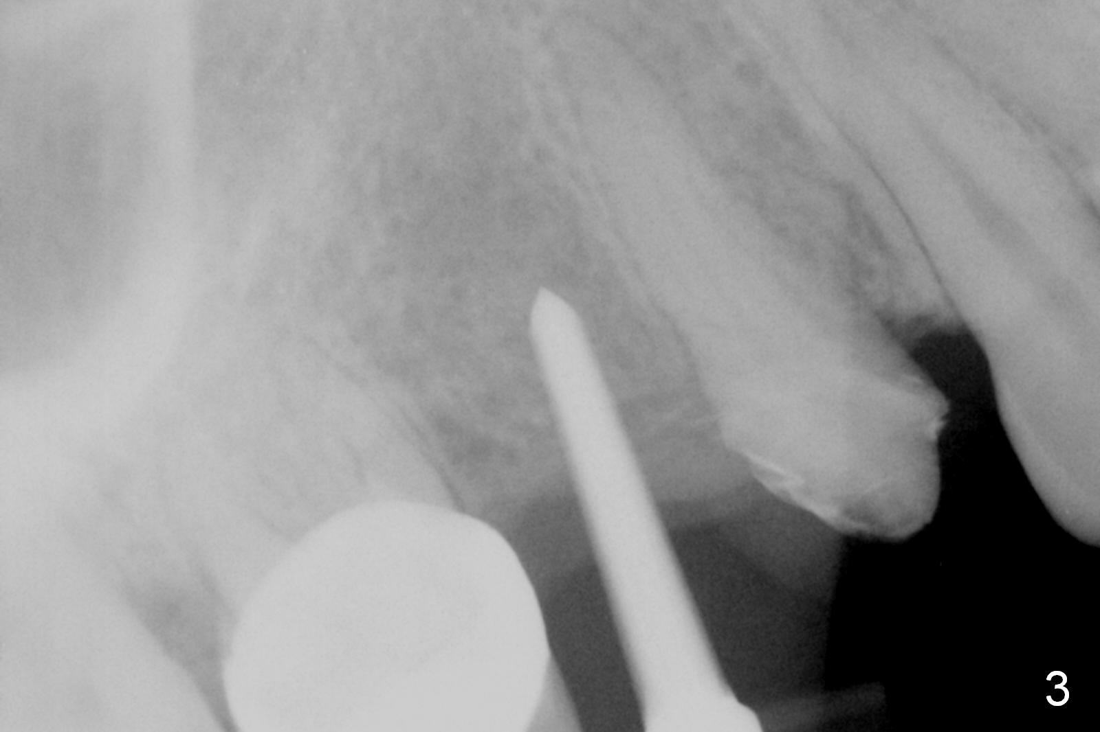
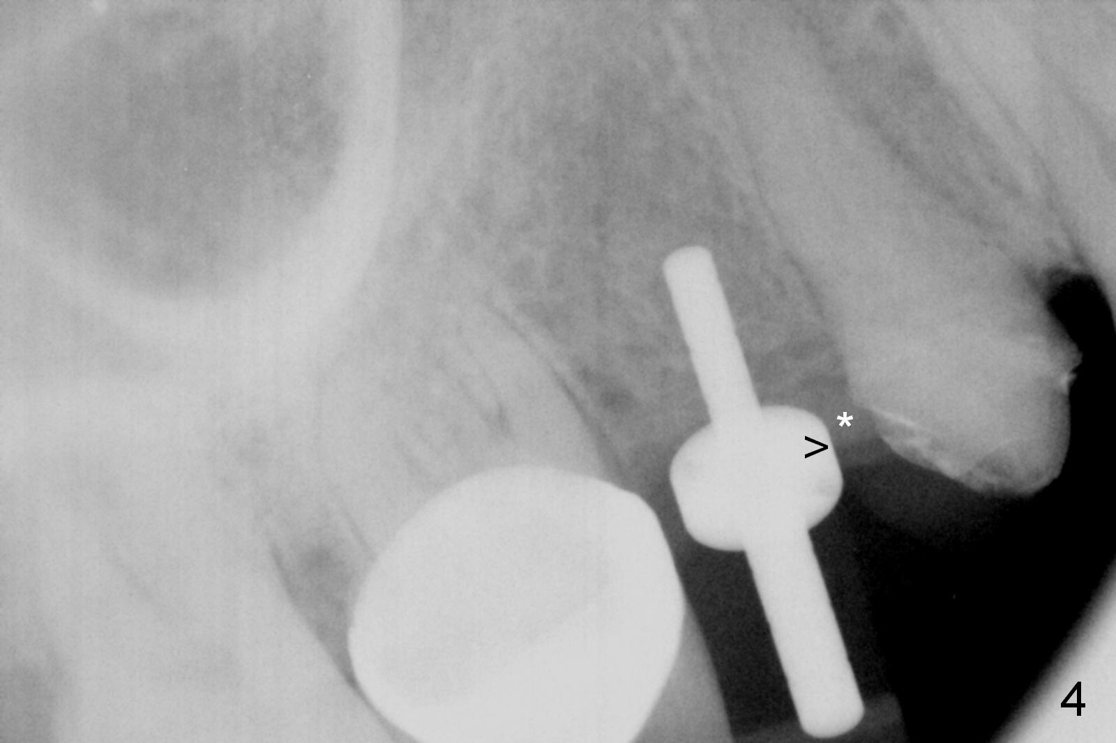
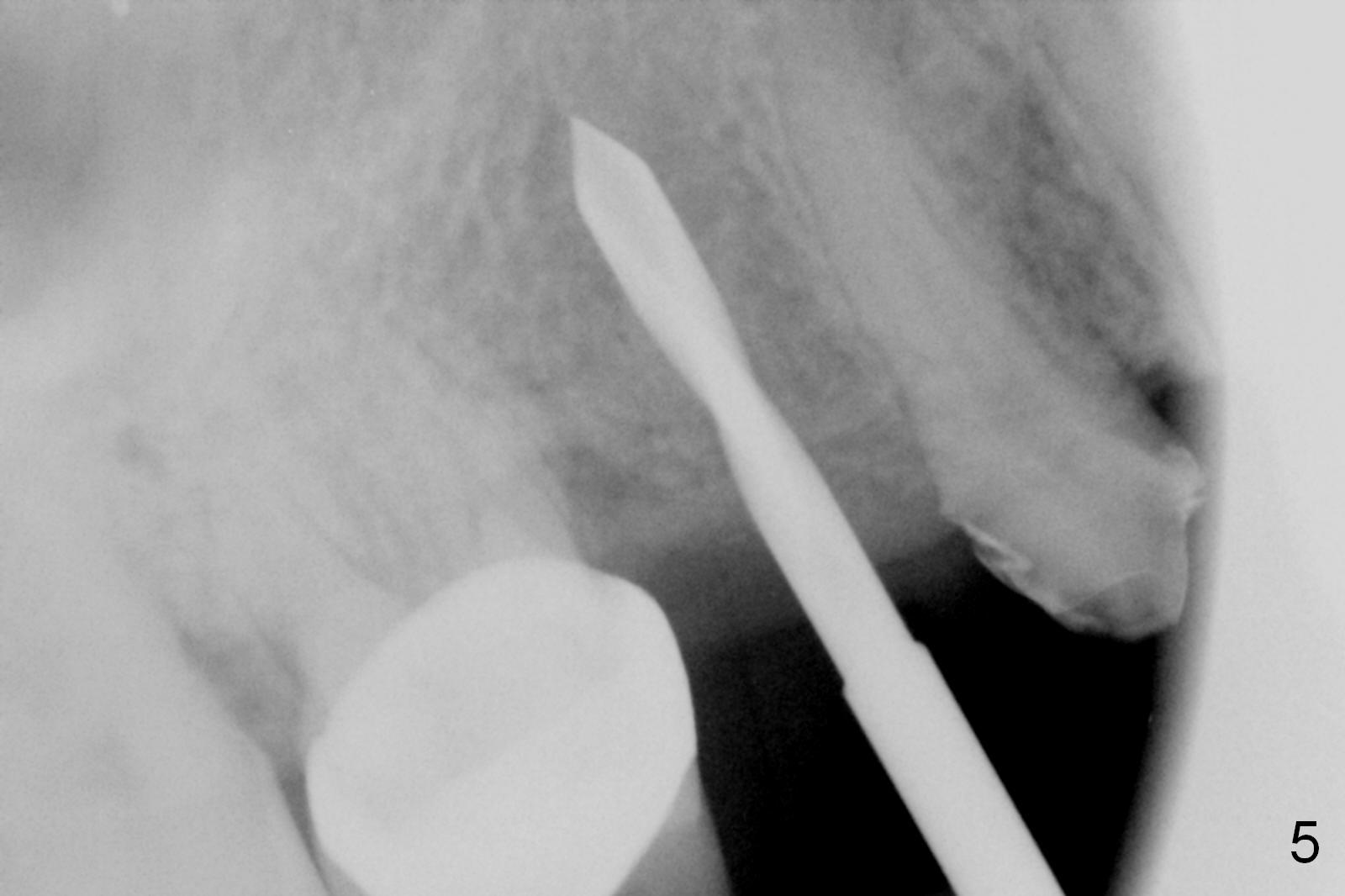
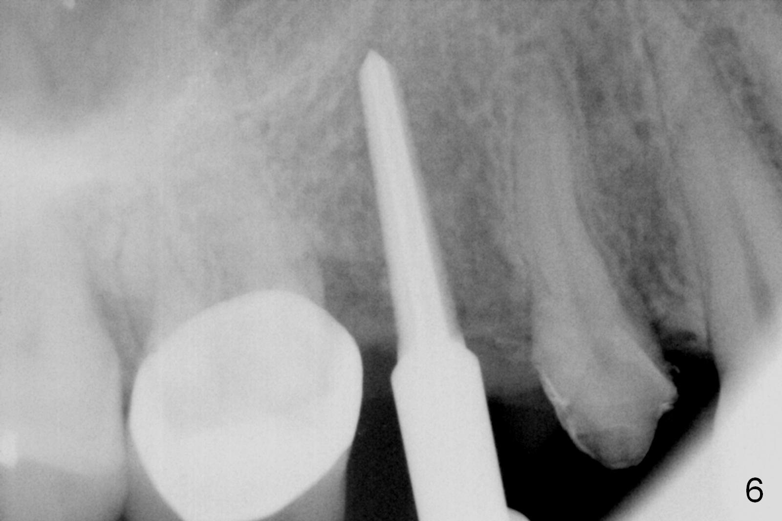
The gingiva is healthy 17 days post pontic removal (Fig.1,2). After making a small incision, #15 blade is used to start bone expansion, followed by Magic Split for ~ 8 mm (gingival level, Fig.3). The trajectory is corrected with using IBS pilot drill, but is not shown with insertion of a Guide Pin (Fig.4). The large portion of the latter (>) is blocked by the provisional of the neighboring tooth (*). The entrance of the osteotomy is moved distally with a Lindamann bur, followed by a 2 mm drill (Fig.5). Then bone expansion is much easier with Magic Expander (ME) 30 at 15 mm (Fig.6). Following ME38 half the depth mentioned last, a 4x13 mm implant is placed with insertion torque >35 Ncm (Fig.7). Pair abutment (4x4(3) mm) is placed for an immediate provisional.
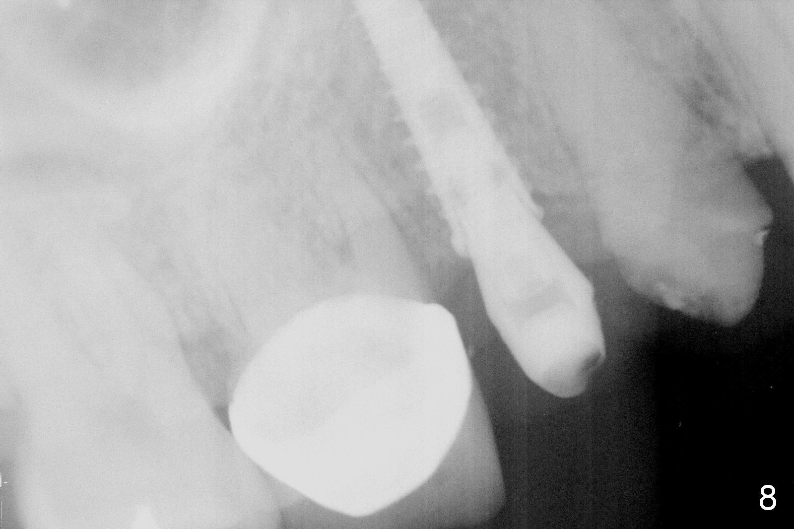
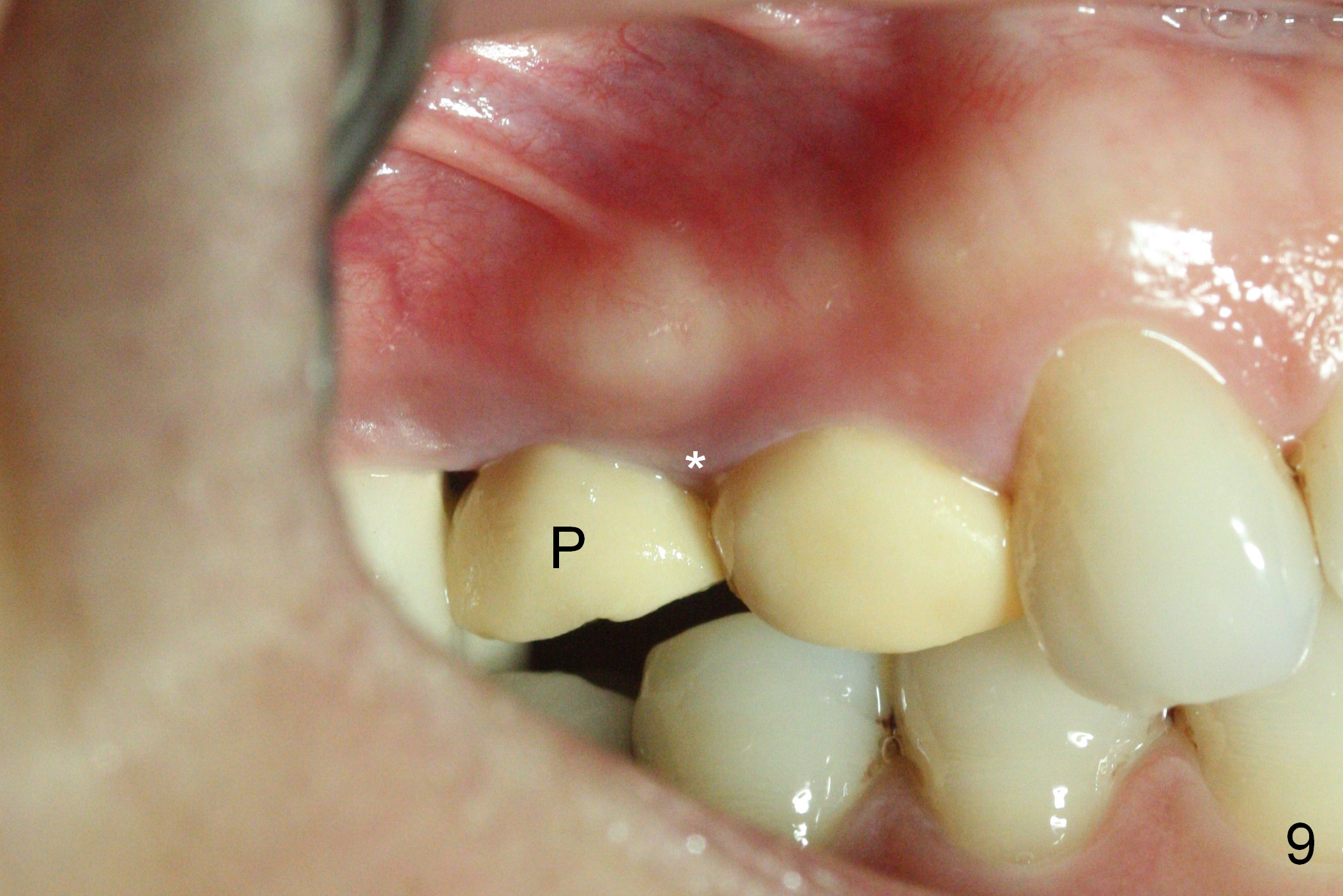
The patient returns for impression 4 months postop. There is no crestal bone resorption (Fig.8). The provisional (Fig.9 P) maintains the mesial papilla (*).
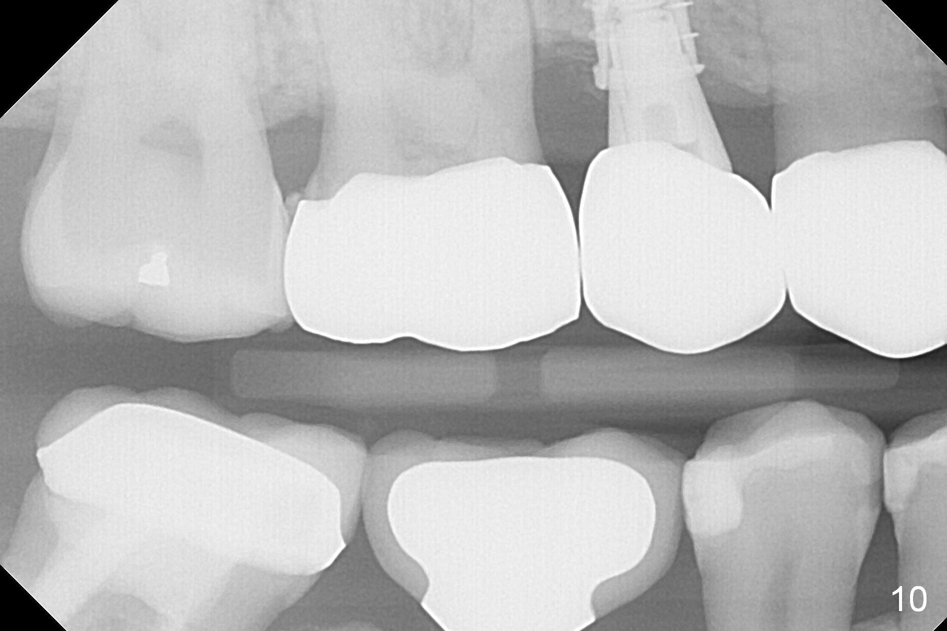
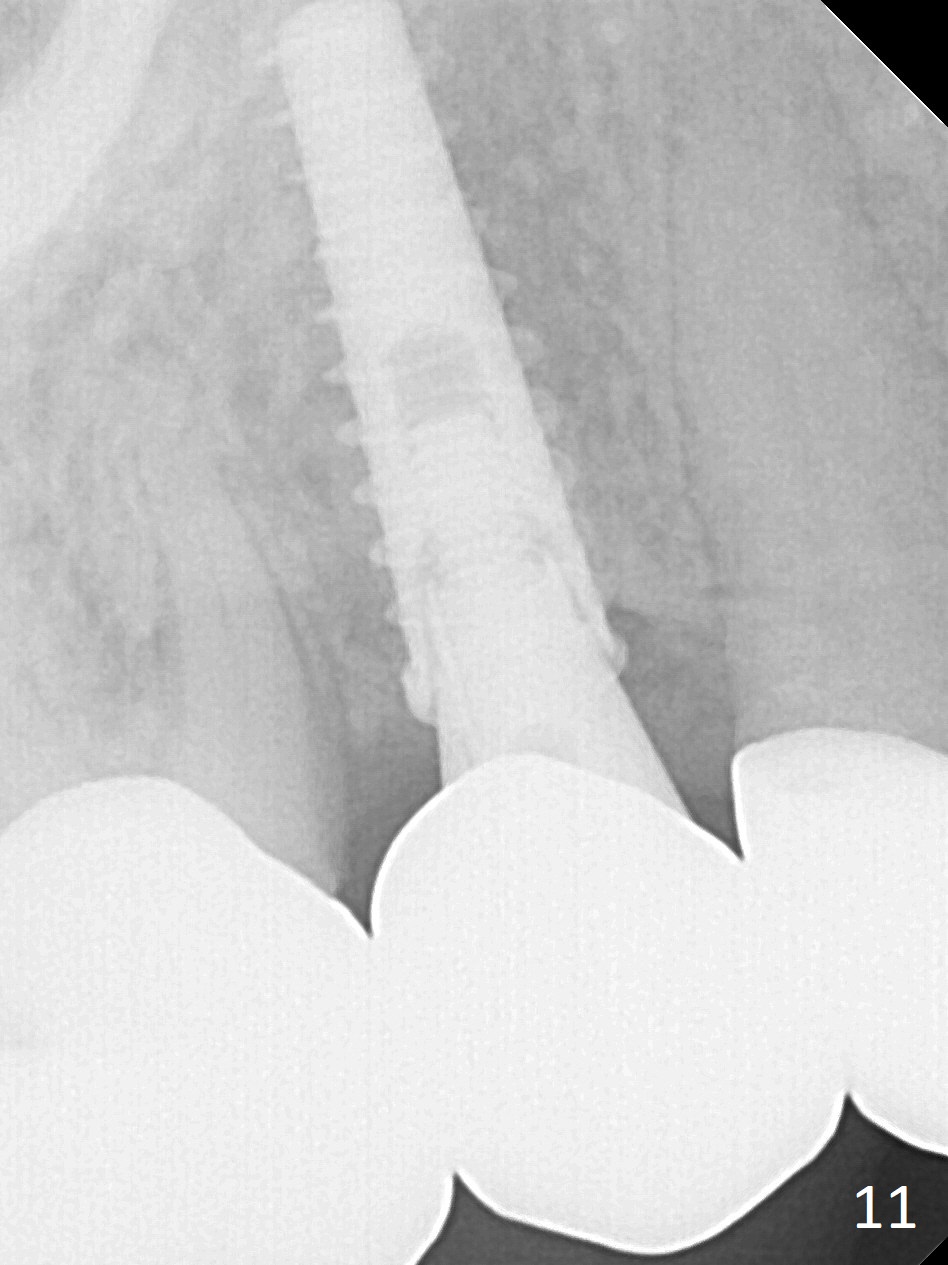
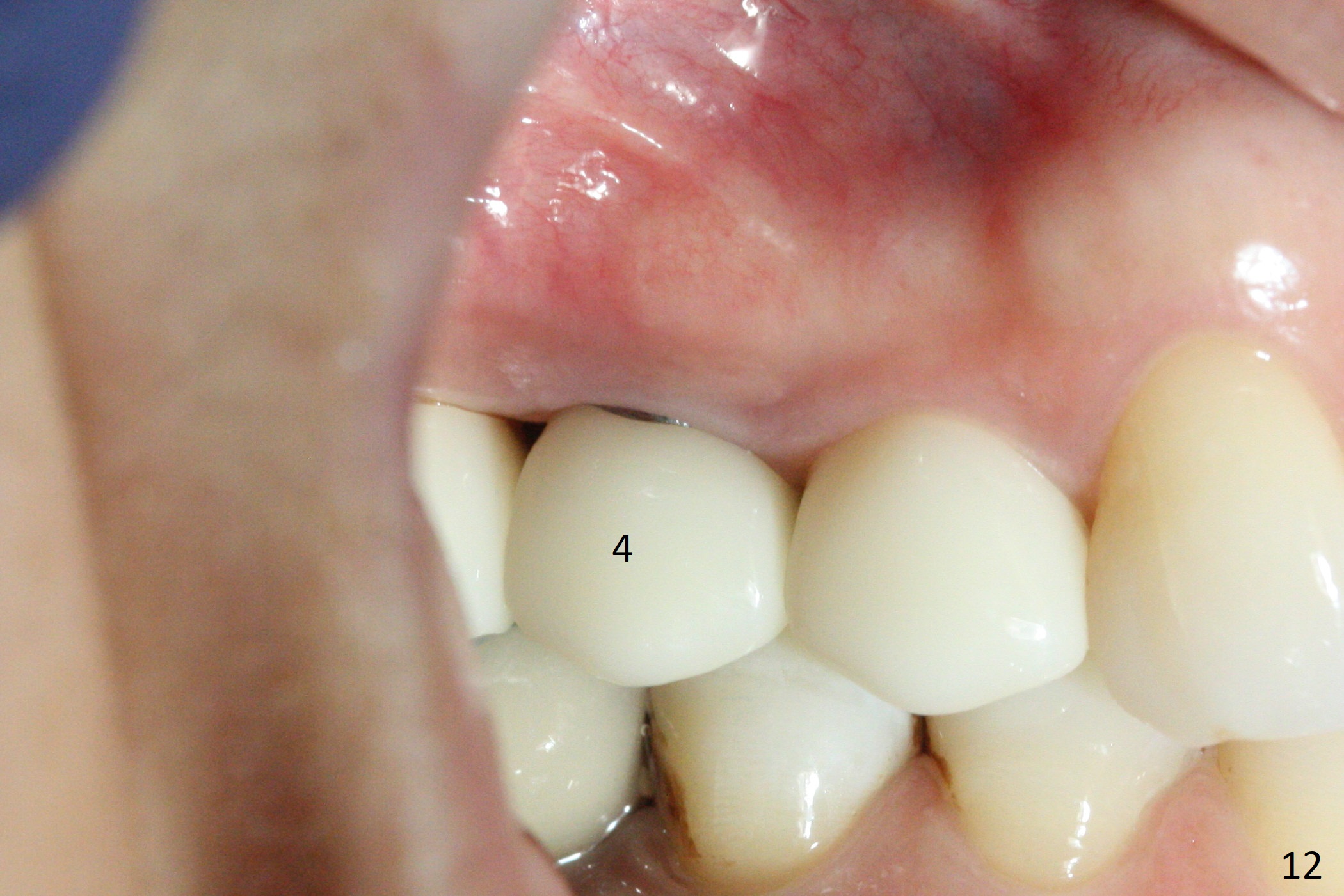
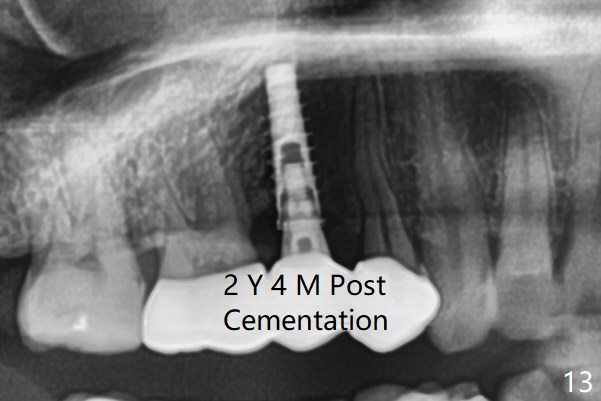
Crowns at #3-5 are cemented 6 months postop (Fig.10). No bone resorption is observed 4.5 months post cementation (Fig.11). The soft tissue is healthy 10 months post cementation (Fig.12). The abutment remains incompletely seated, although the crown is not loose 2 years 4 months post cementation (Fig.13).
Return to Upper Bicuspid Immediate Implant, Posterior Immediate Provisional, IBS, Parallel Pin
Xin Wei, DDS, PhD, MS 1st edition 07/29/2016, last revision 03/28/2021