.jpg)

|
|
|
|
|
|
|
|
|
|
|
|
|
|
|
|||
Immediate Impression
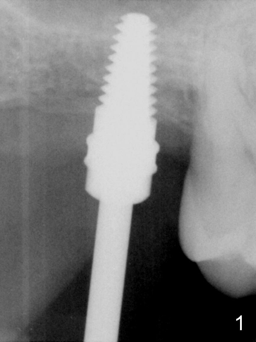
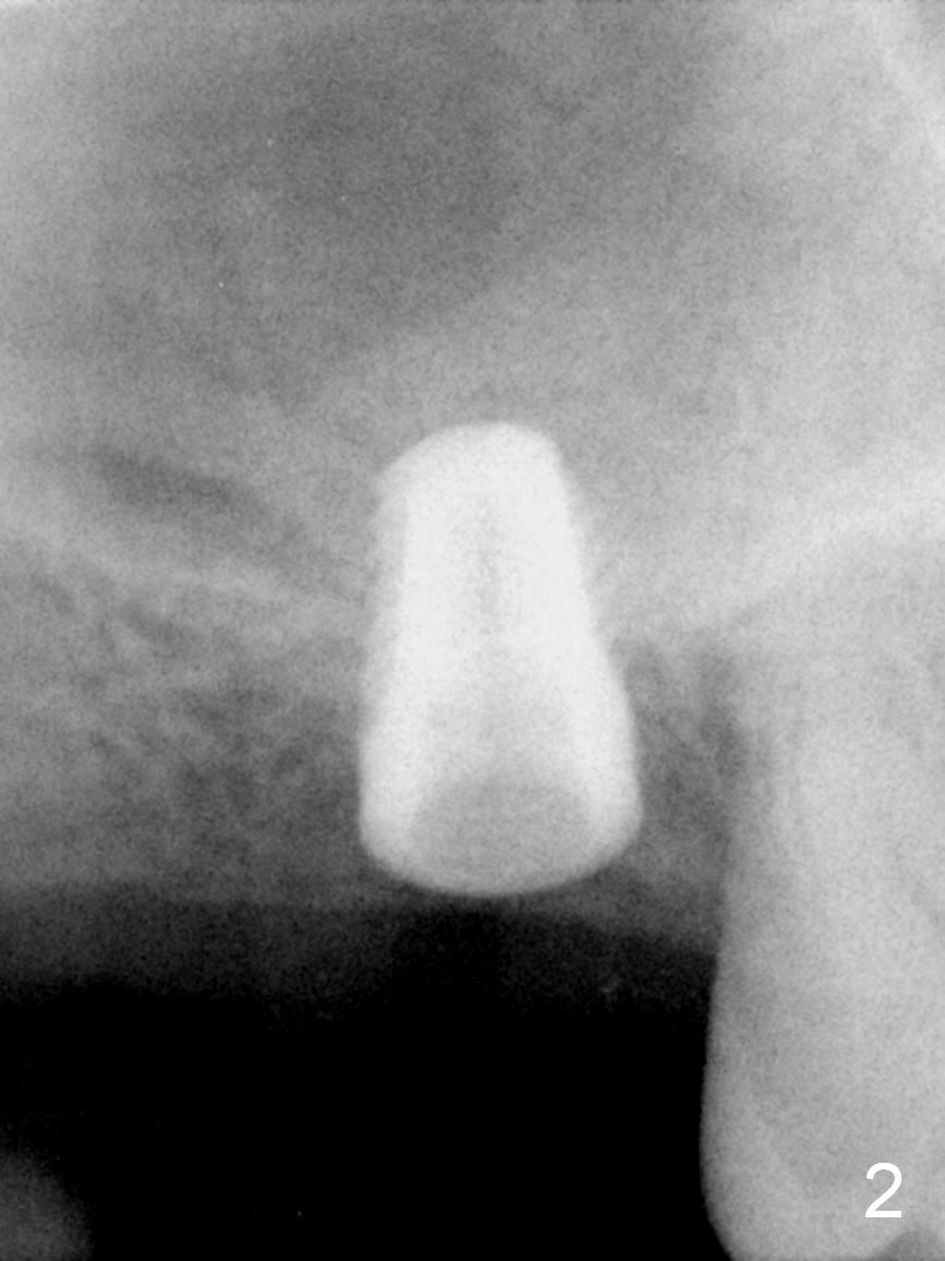
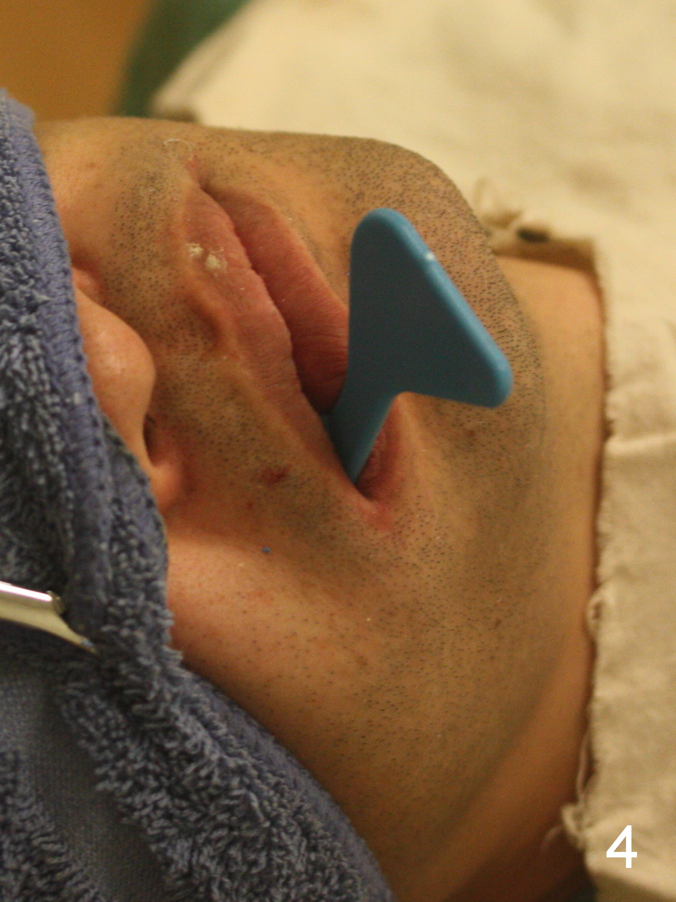
The ridge at the site of #2 is mild to moderately atrophic. While a straight incision ~ 9 mm long is being made over the ridge, the underlying bone feels soft. The #15 blade is tapped in to start bone expansion, followed by RT 2-4 and 4.5x11 mm tap at 8 mm at gingival level (Fig.1 T). Allograft and Osteogen is placed in the osteotomy and pushed upward by re-inserting the 4.5x11 mm tap. More bone graft is pushed superiorly by inserting 5x11 mm tap. A 5.3x8 mm bone-level implant is placed with insertion torque of >50 Ncm with sign of sinus lift (Fig.2). A 6.8x4(3) mm cemented abutment is hand tightened (Fig.3). After definitively tightening the abutment of the opposing implant at 35 Ncm, final impression is taken (Fig.4).
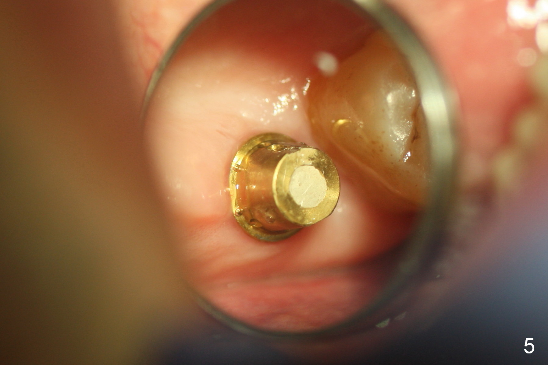
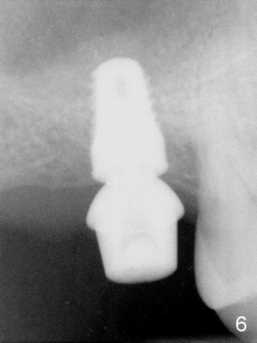
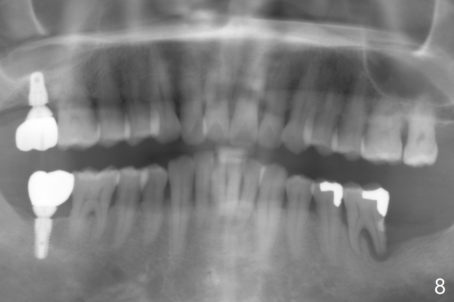
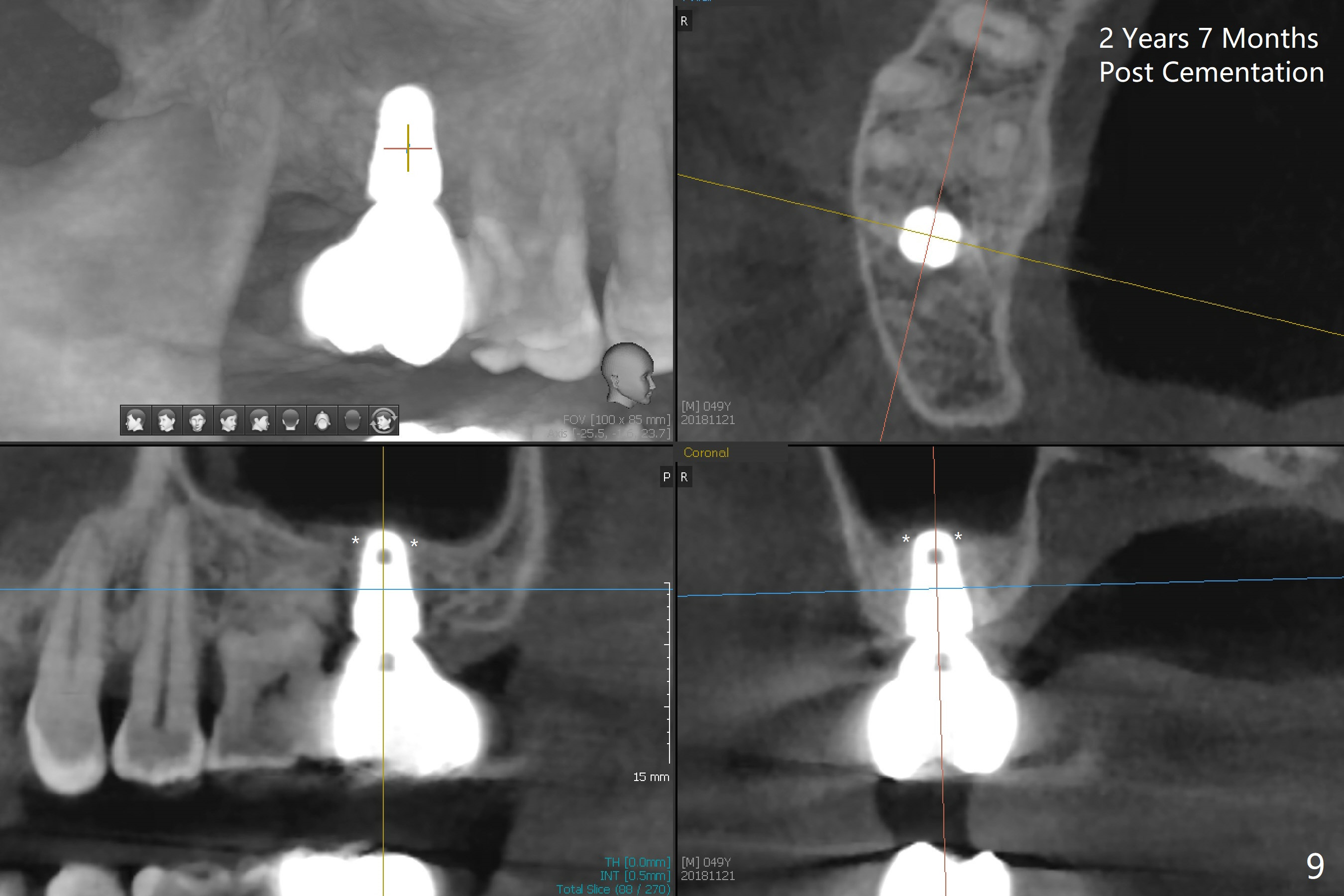
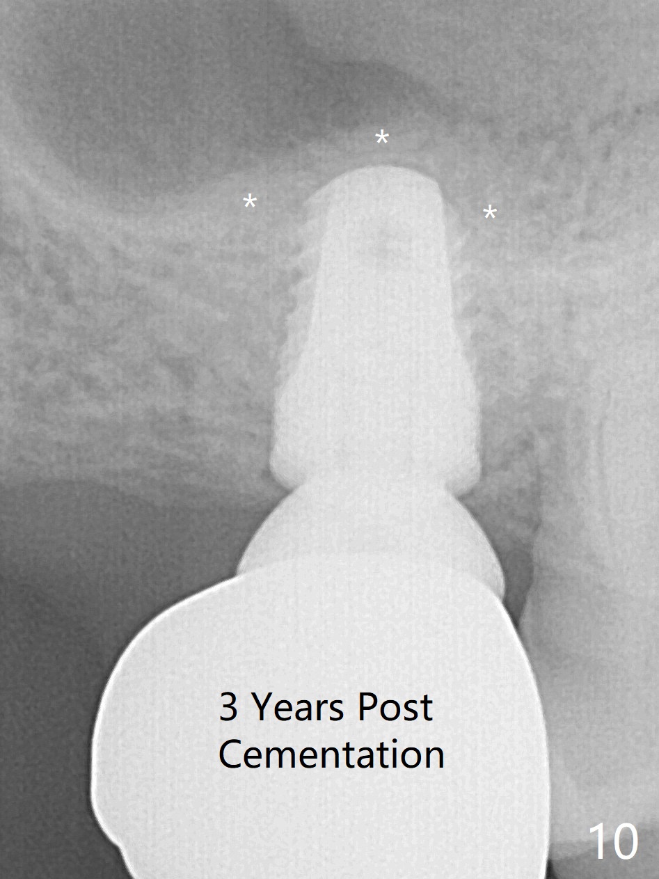
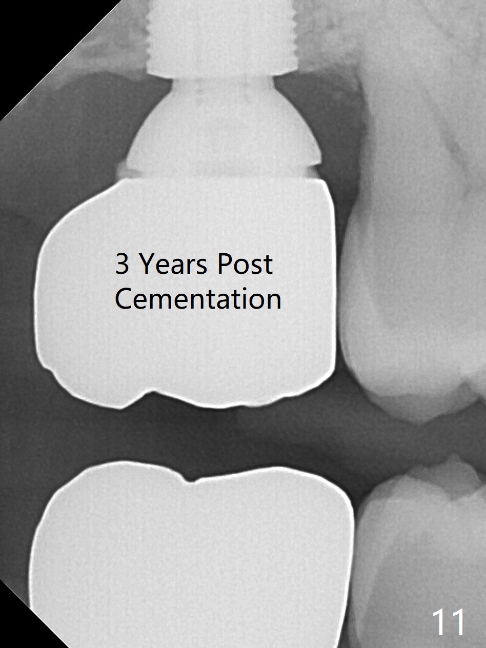
Twenty days postop, the incision heals around the abutment (Fig.5). The provisional stays in place until 3 months postop (Fig.6). The gingiva remains healthy with the supragingival margin of the abutment (Fig.7 ^) when the definitive restoration is tried in (C). In spite of poor crown/implant ratio, there is no bone loss at #2 18 months post cementation, as compared to #15 (Fig.8). CT taken 2 years 7 months post cementation shows that the apex of the implant is covered by the bone (Fig.9 *). Sinus lift remains distinct (Fig.10 *) and there is no crestal bone loss (Fig.11) 3 years post cementation.
Return to Upper Molar Immediate Implant, Immediate Posterior Provisional, 19 Xin Wei, DDS, PhD, MS 1st edition 12/29/2015, last revision 04/24/2019