|
|
|
|
|
|
Second Treatment Plan for an Upper 2nd Molar Immediate Implant
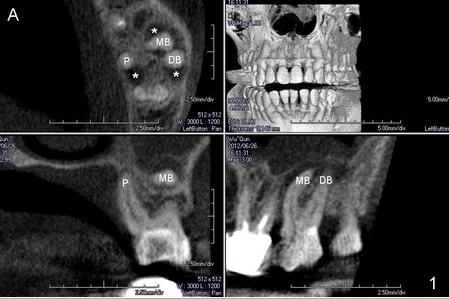
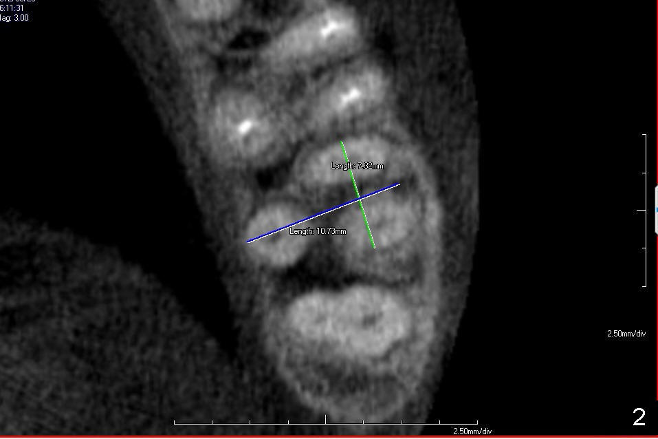
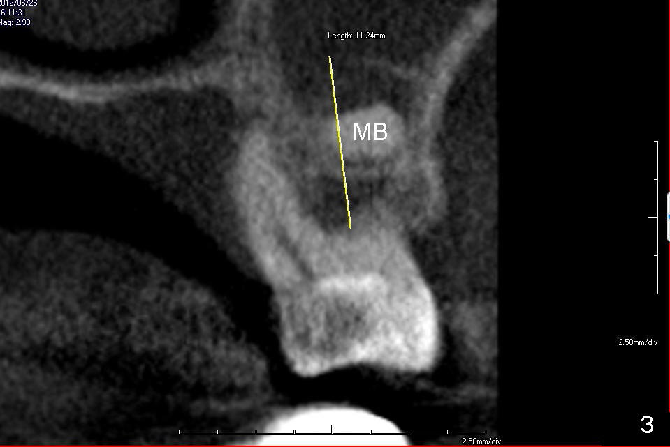
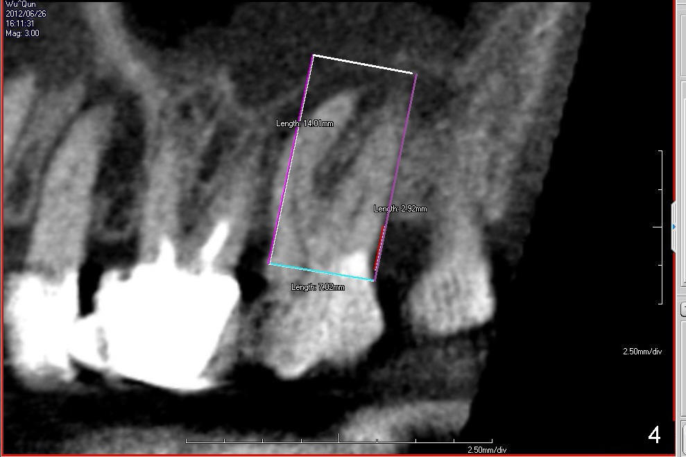
If the septum of the tooth #15 is destroyed by the infection, a larger implant is used instead (using socket walls for stability), tapered or cylindrical, 6 or 7x14 mm (Fig.3), depending upon tap stability. In Fig.1A, the axial section is made at the apices of the three roots. It shows that there is a lesion around each apex (*). Before implantation, bone graft is to be placed using a small graft syringe. Fig.2 axial cut is made in the middle level of the septum, attention being paid to the widths of the roots, the largest dimension (diameter) of an implant to be used. Fig.3 measures the height of the septum.
Although CBCT is helpful, bone quality can be only decided upon tooth
extraction, i.e., while using a drill or osteotome.
Xin Wei, DDS, PhD, MS 1st edition 02/16/2014, last revision 02/16/2014