
|
|
|
|
|
|
|
|
|
|
|
|
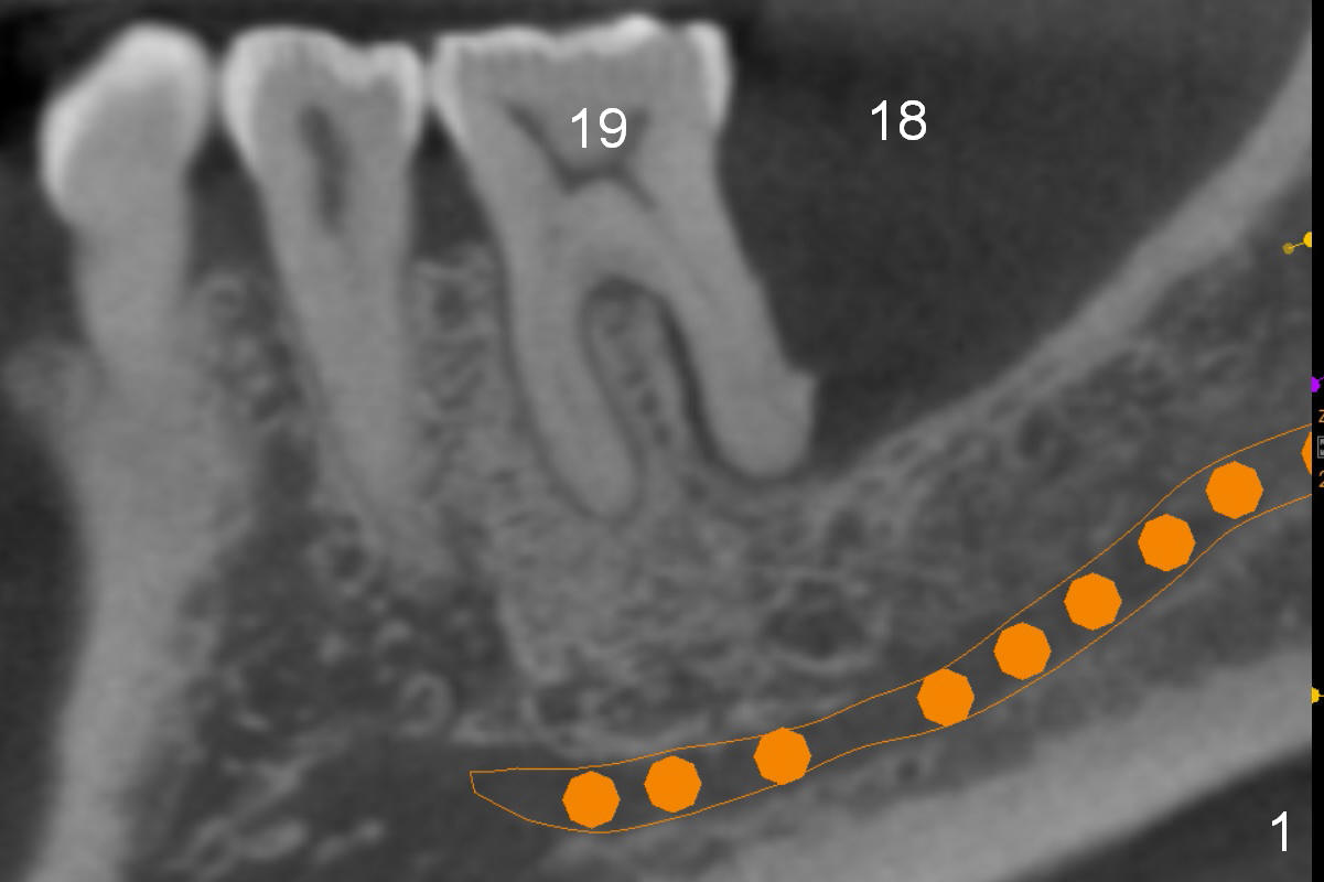
Parallel Pins
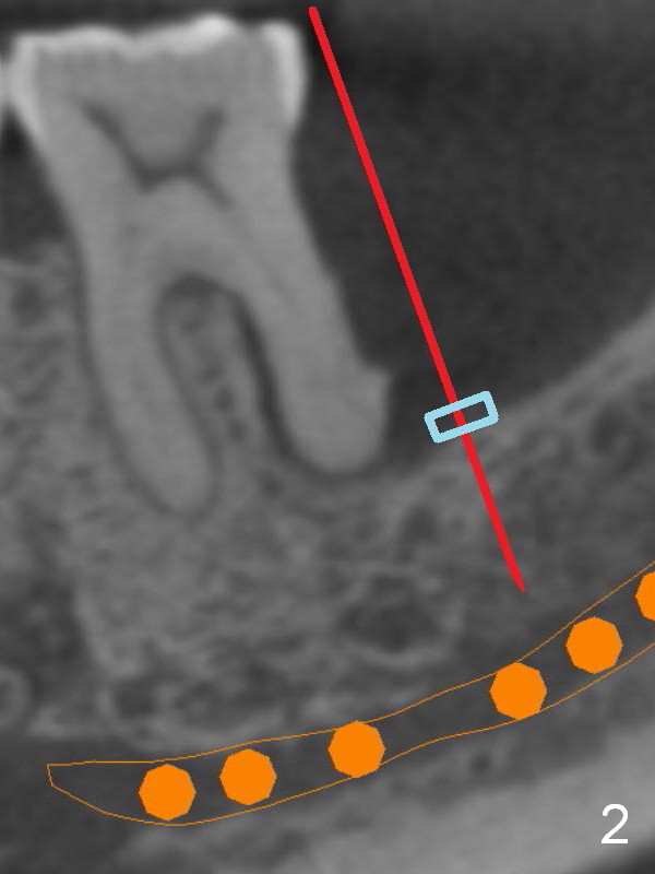
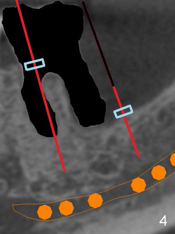
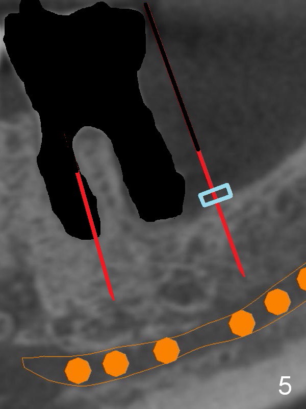
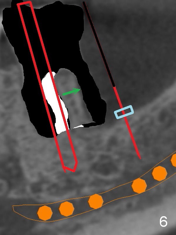
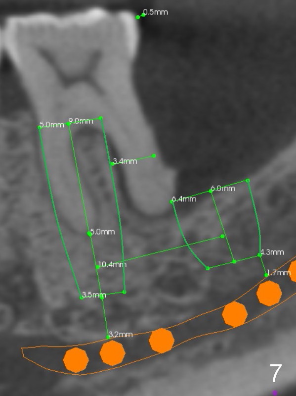
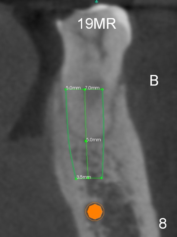
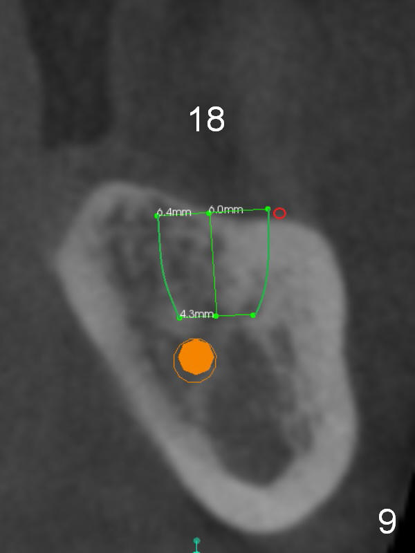
There is severe bone resorption around the distal root of the lower left 1st molar of the 56-year-old man (WG), while the loss of the 2nd molar is associated with limited bone height (Fig.1). A 5x14 mm implant will be placed at the mesial socket of #19 and 5.9 or 6.4x6 mm one is at the mesial site of #18 (Fig.7-9). Use a 2 mm pilot drill with 6 mm stopper from Sinus Master Kit (with extension) to initiate osteotomy at #18 immediately distal to the crown of #19, parallel to the long axis of the latter (Fig.2). After inserting a parallel pin at #18, extract the tooth #19 (Fig.3, antibiotic pending) and start osteotomy with a 2 mm pilot drill with 14 mm stopper (Fig.4). Insert the calibrated parallel pin at #19 (Fig.5) and measure the distance between the two parallel pins (approximately 10 mm, Fig.7). Sequential osteotomy and application of the Tatum taps (Fig.6 red rectangle) will push the septum distal (green arrow). As to #18 osteotomy, trephine, final and tap drills are to be used with control of the depth: 6 mm (Fig.7,9).
Return to Upper Arch, Lower Molar Immediate Implant
Xin Wei, DDS, PhD, MS 1st edition 01/24/2016, last revision 01/24/2016