|
|
|
|
|
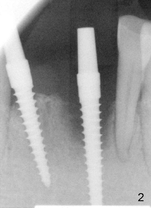
The teeth #24 and #26 are extracted and replaced by 3x20 and 3x17 mm one piece implants, respectively (Fig.2). It is difficult to initiate osteotomy at the site of #24 because of severe bone loss and deep socket. Incisions are made for direct vision.
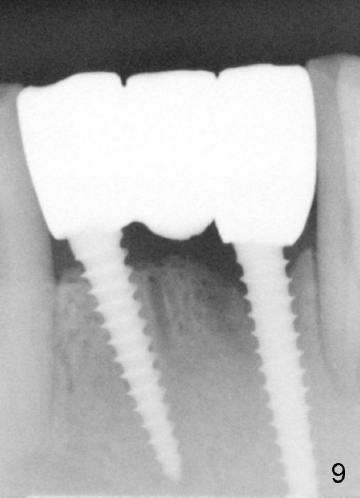
Fig.9 shows apparent bone growth around #24 implant (at the crest) 6,9 months post cementation and surgery, respectively.
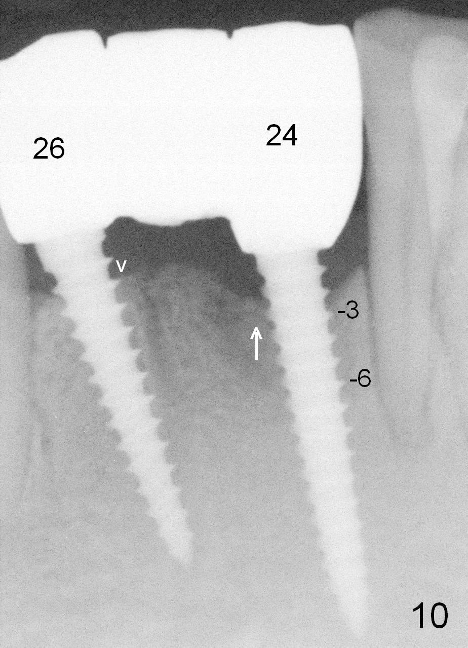
Fig.10 shows that bone grows coronally (arrow) to cover 3 threads (from #6 thread to #3) at the site of #24 one year after functioning.
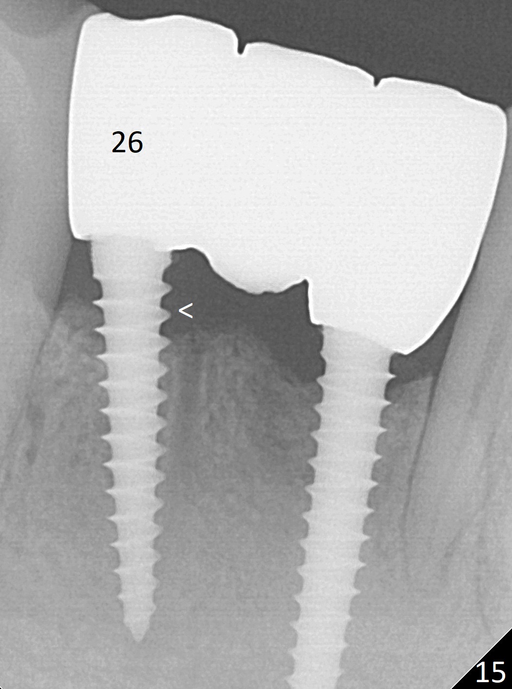
In fact, periimplantitis develops at #26 two years of absence of perio maintenance (because of atrial fibrillation with blood thinner; Fig.15, 3 years 7 months post cementation). The implant at #26 should have been smaller and placed deeper (Fig.2).
Return to Incision Dehiscence Last Next
Xin Wei, DDS, PhD, MS 1st edition 03/24/2013, last revision 12/16/2018