.jpg)
|
|
|
|
|
|
|
|
|
|
||
|
|
|
|
||
Double Check Bone Graft
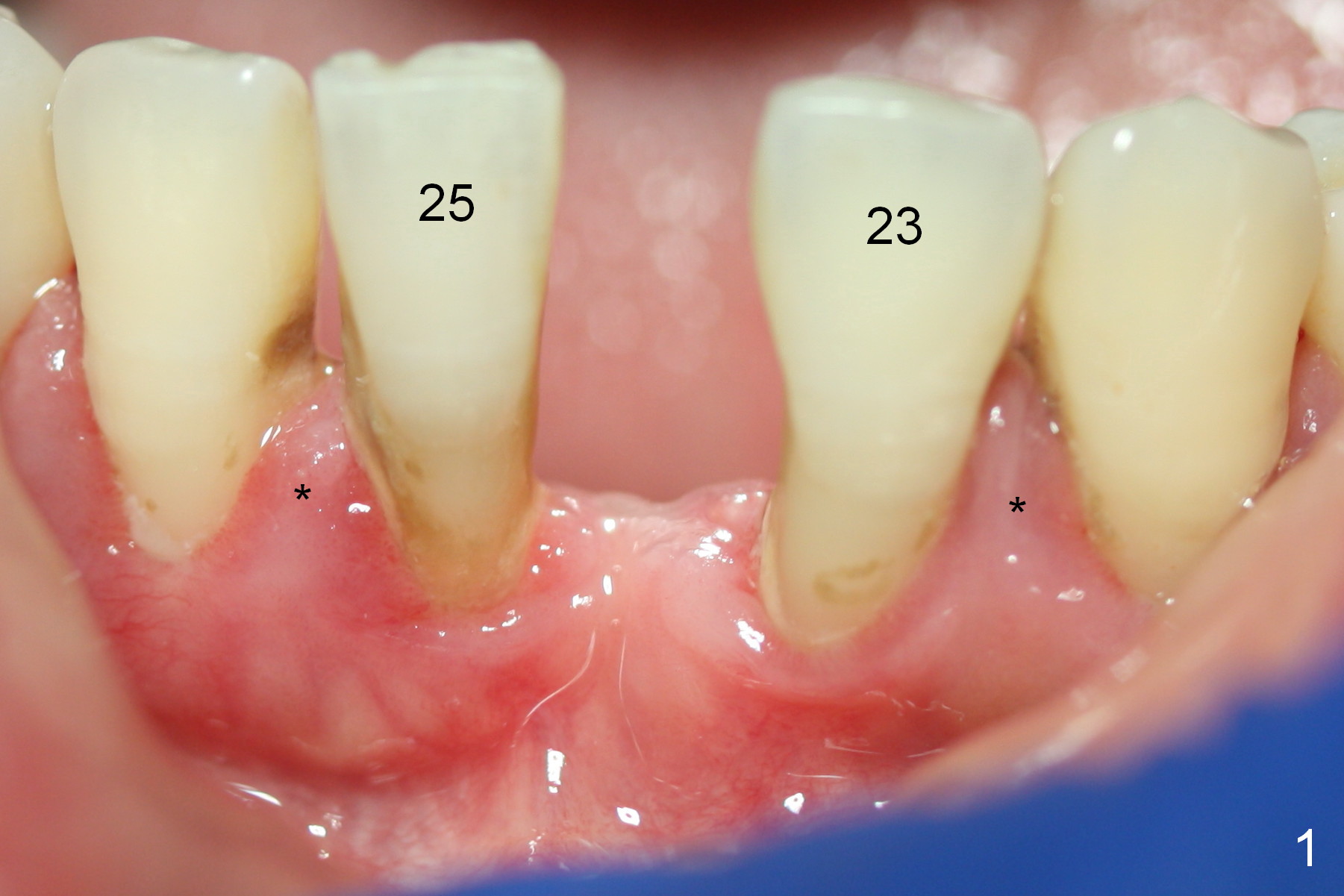
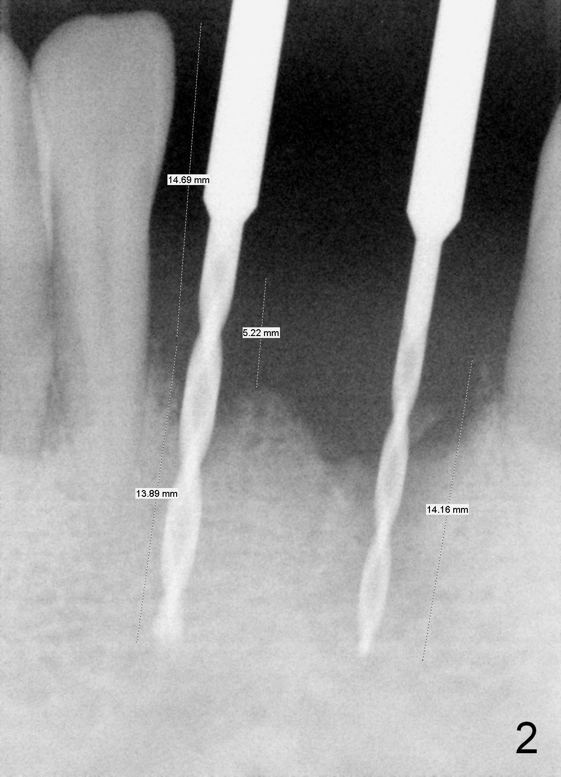
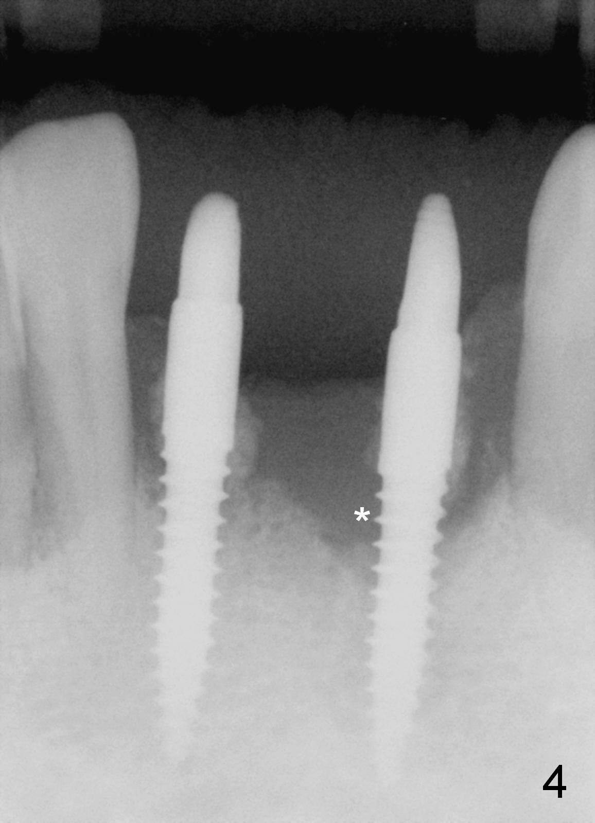
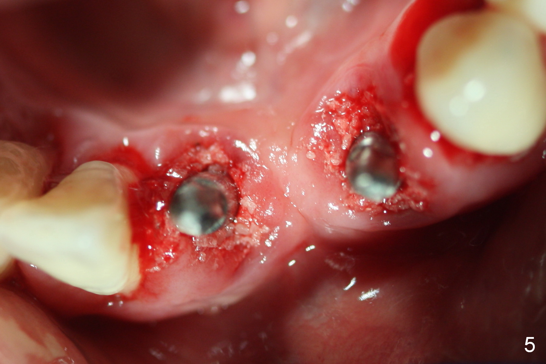
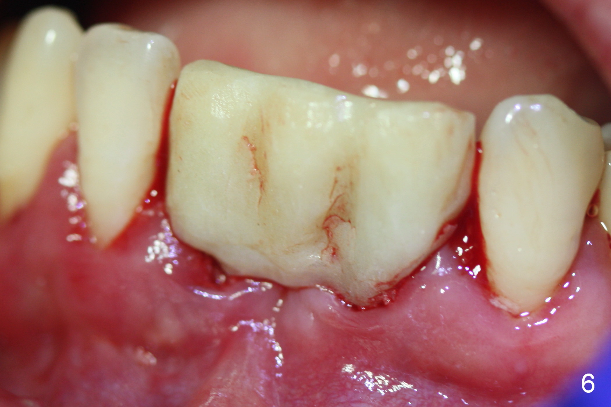
When the patient returns for implant placement, the tooth #23 turns out to be as severely periodontally involved as the tooth #25 (Fig.1). After extraction, osteotomy starts at these sites. It appears that the future crown/root ratio will be 1:1 (Fig.2); the gingiva is 5 mm thick. Therefore 2 of 3x14 mm 1 piece implant are placed with 4 mm gingival cuff (Fig.3 insertion torque >60 Ncm). After preparation of the abutments and packing bone graft, a new PA is taken (Fig.4,5). It appears that the graft is lacking mesial to the implant at the site of #23 (Fig.4 *). More bone graft is then added to the area, which will facilitate bone healing. Finally an immediate provisional bridge is fabricated (Fig.6).
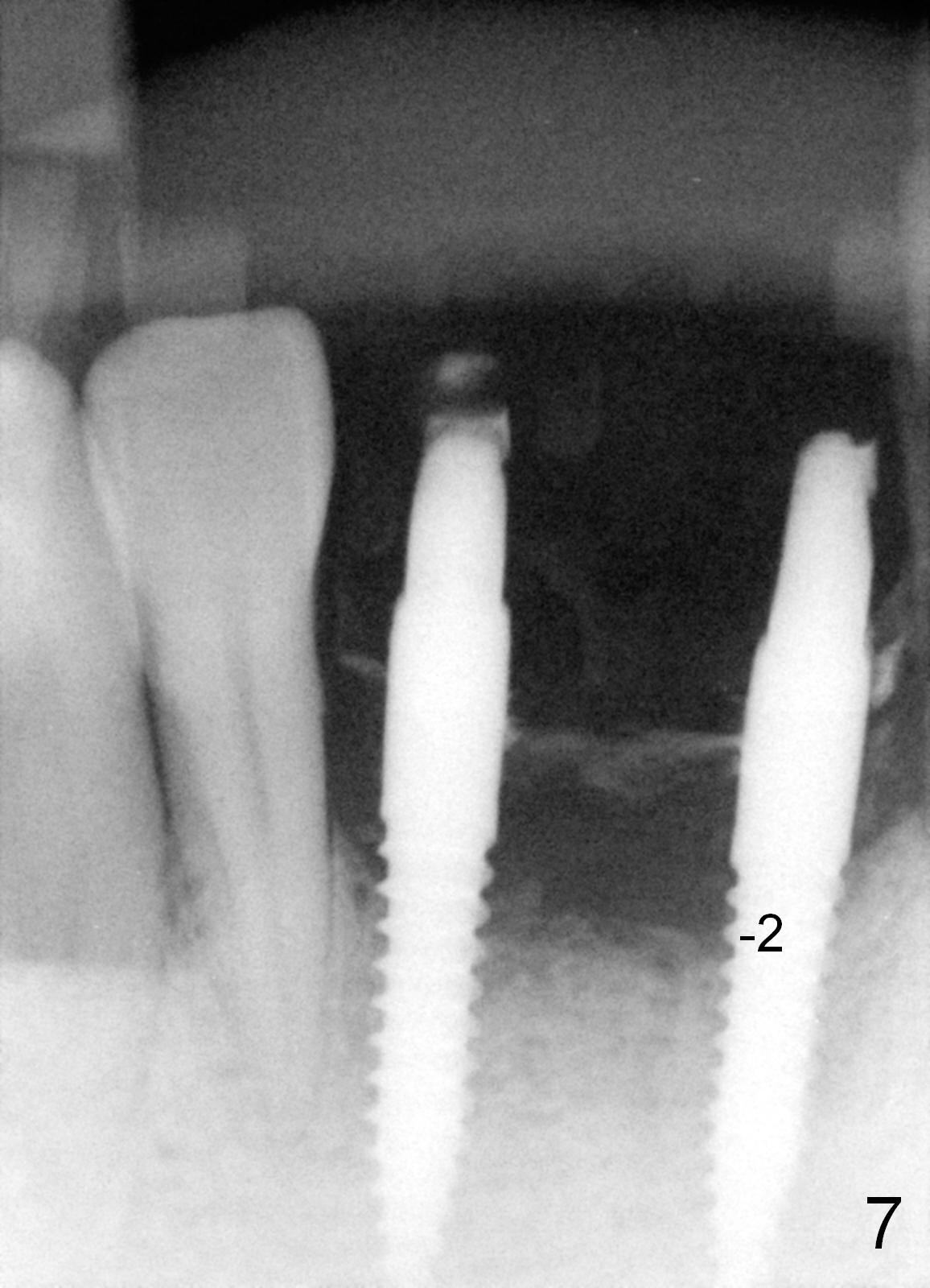
The patient returns 4 months postop, wearing the same provisional. The gingiva around the implants and neighboring teeth is inflamed due to poor oral hygiene and unmodified provisional. Nevertheless, there is bone regrowth at the mesial crest of the implant at #23 (Fig.7).
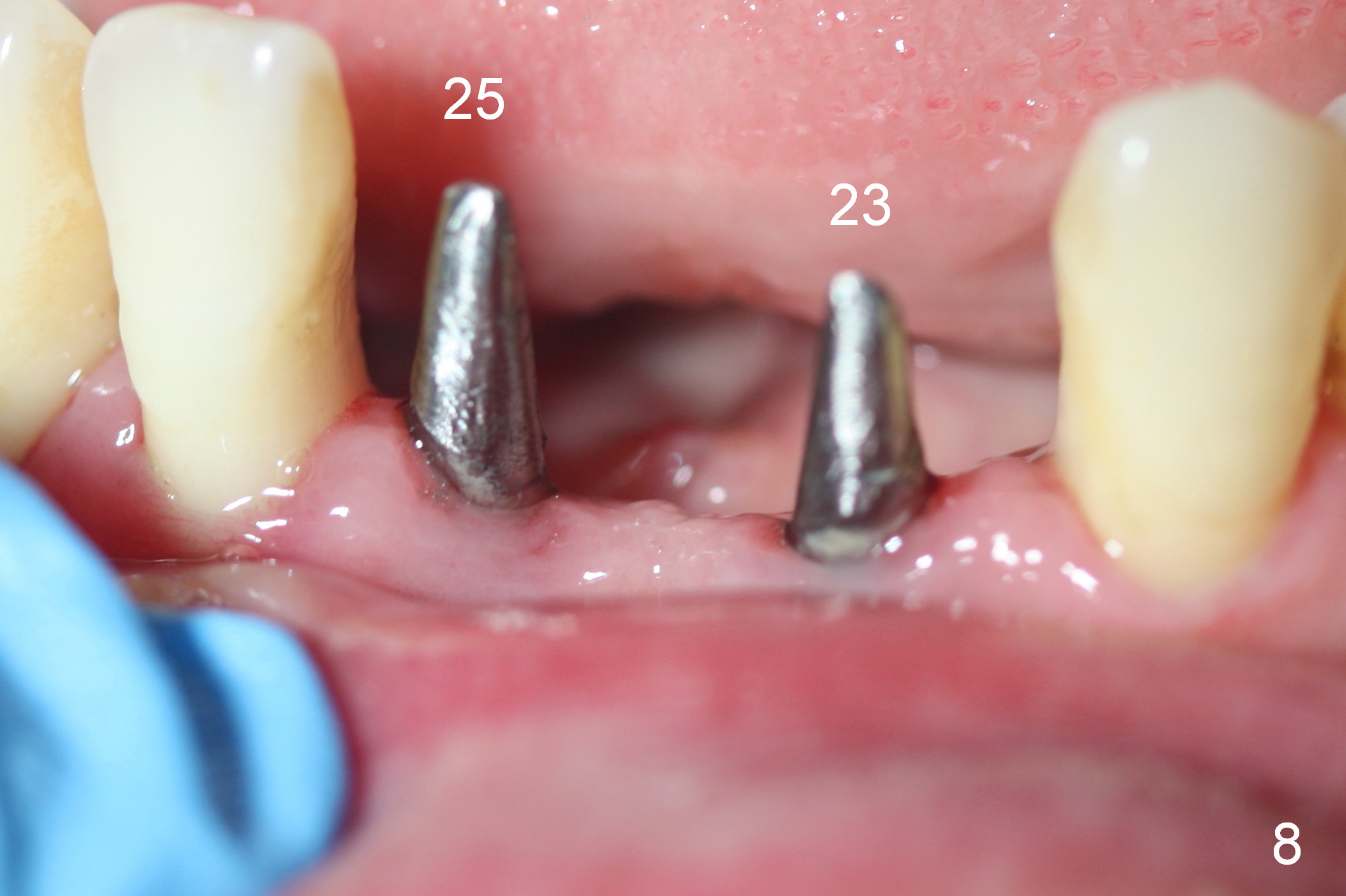
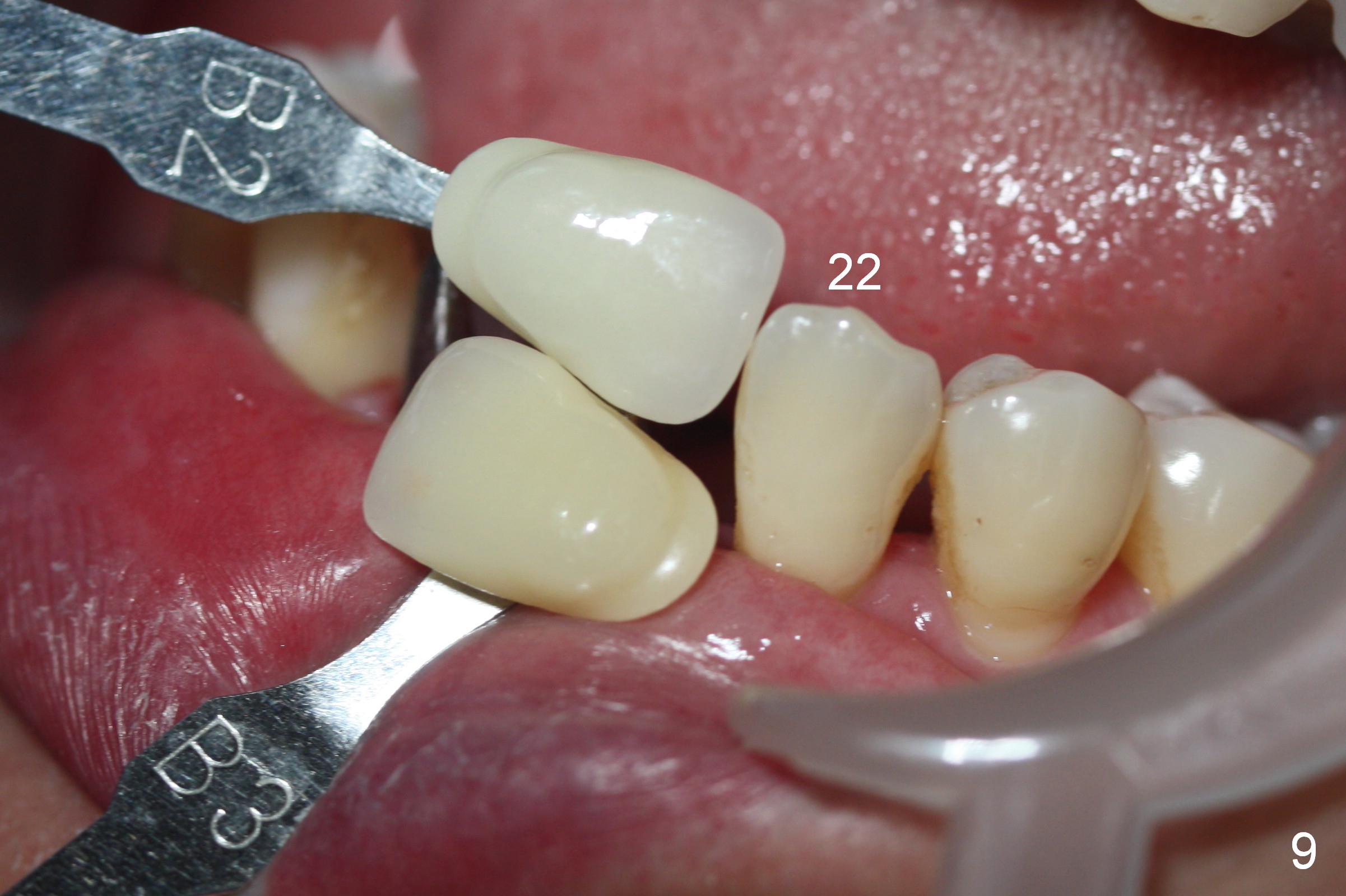
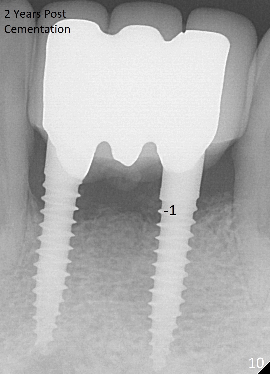
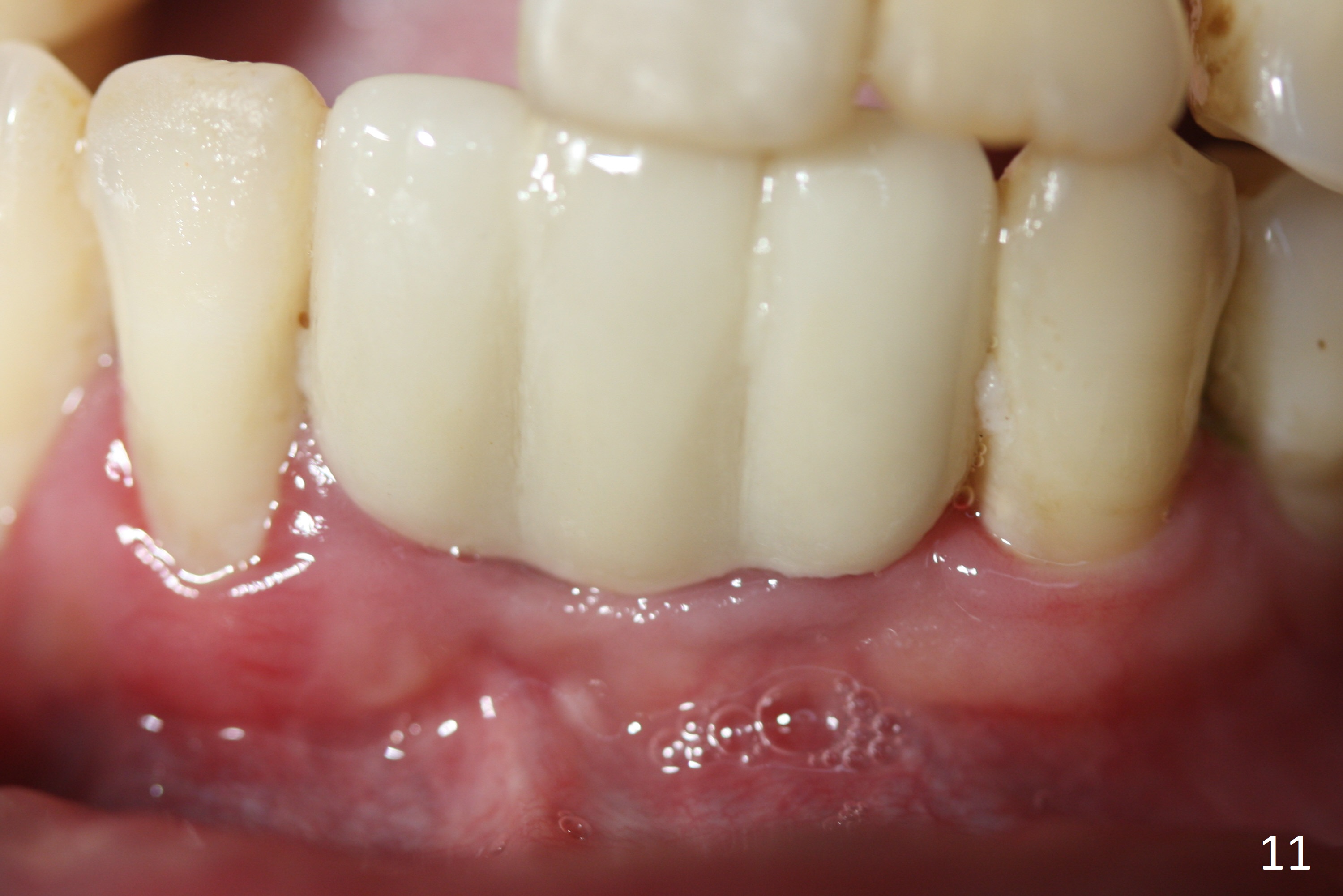
Twenty days later, the gingiva is healthy around the implants (Fig.8). Impression is taken with shade selection (Fig.9). The 1st thread appears to have been covered by the bone 2 years post cementation (Fig.10); the gingiva seems to be healthy (Fig.11).
Return to Lower Incisor Immediate Implant,
Technicians, #7-10
Xin Wei, DDS, PhD, MS 1st edition 05/12/2015, last revision 01/27/2019