|
|
|
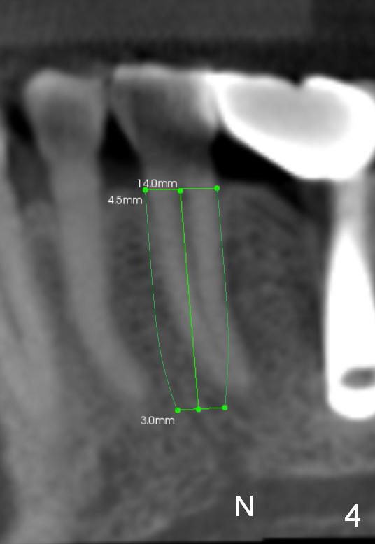
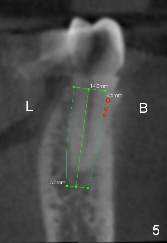
CT sagittal section of a similar case shows that the root is also long, but curved (Fig.4). A 4.5x14 mm implant is slightly longer than the root. The apex of the implant has distance to the underlying nerve (N). The coronal section shows that the implant should be placed lingually (Fig.5 L), since the buccal (B) plate is thin. When the implant is placed, there should be a buccal gap, to be filled with bone graft (red circles).
Xin Wei, DDS, PhD, MS 1st edition 03/17/2015, last revision 03/17/2015