|
|
|
|
|
|
|
|
|
|
|
|
|
|
|
|
|
|||
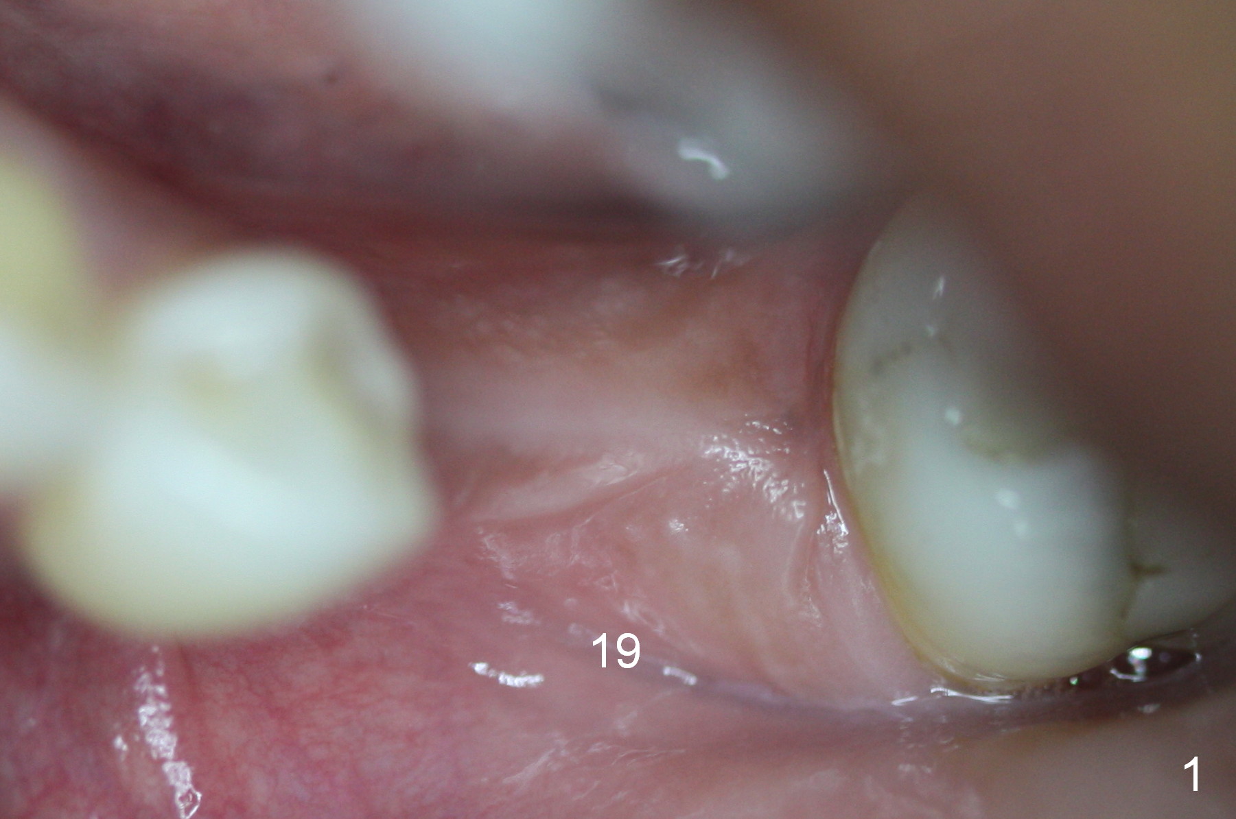
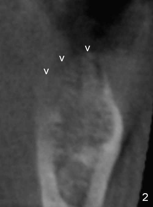
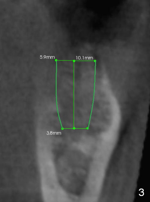
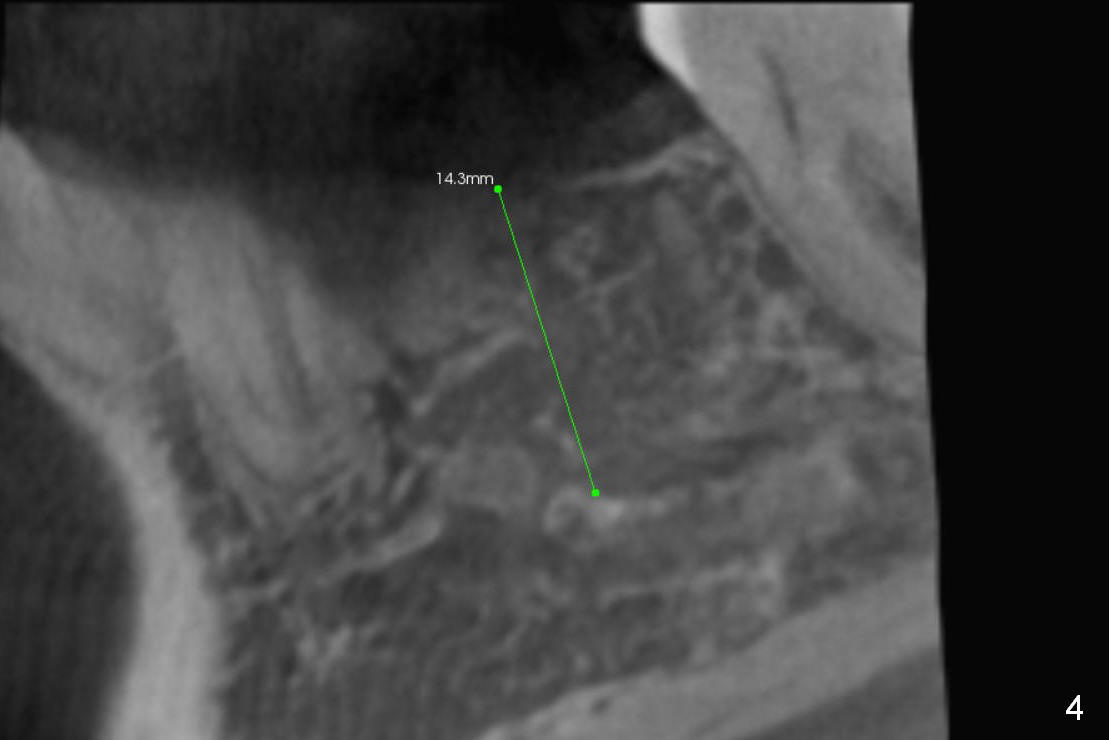
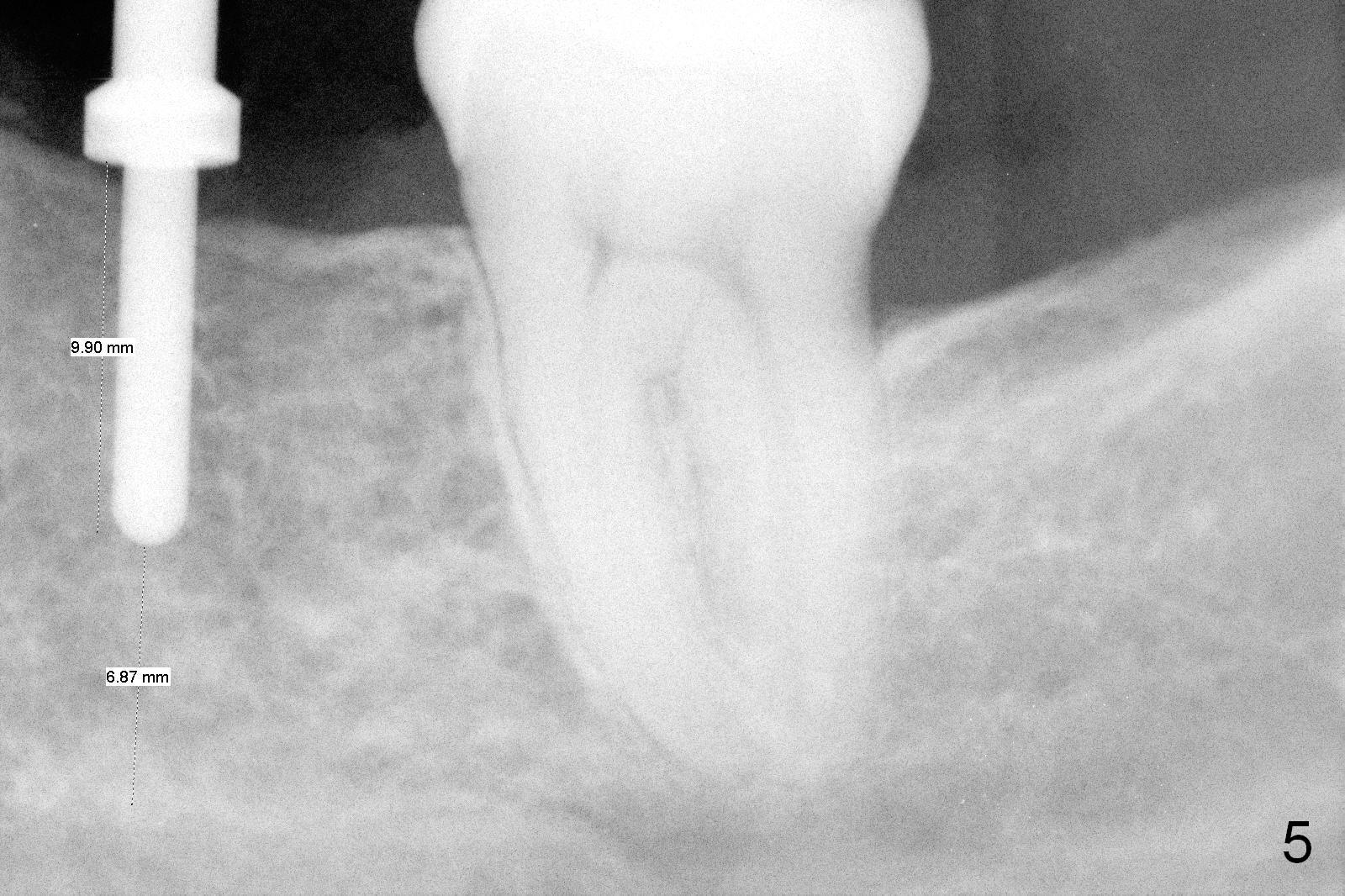
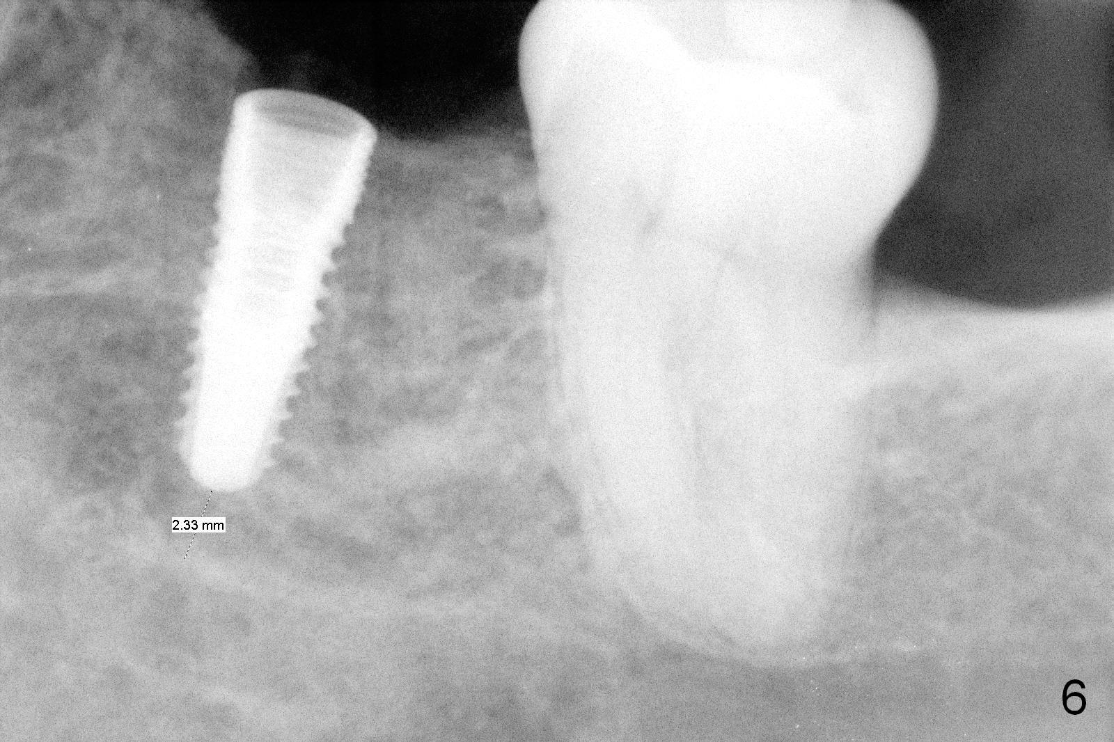
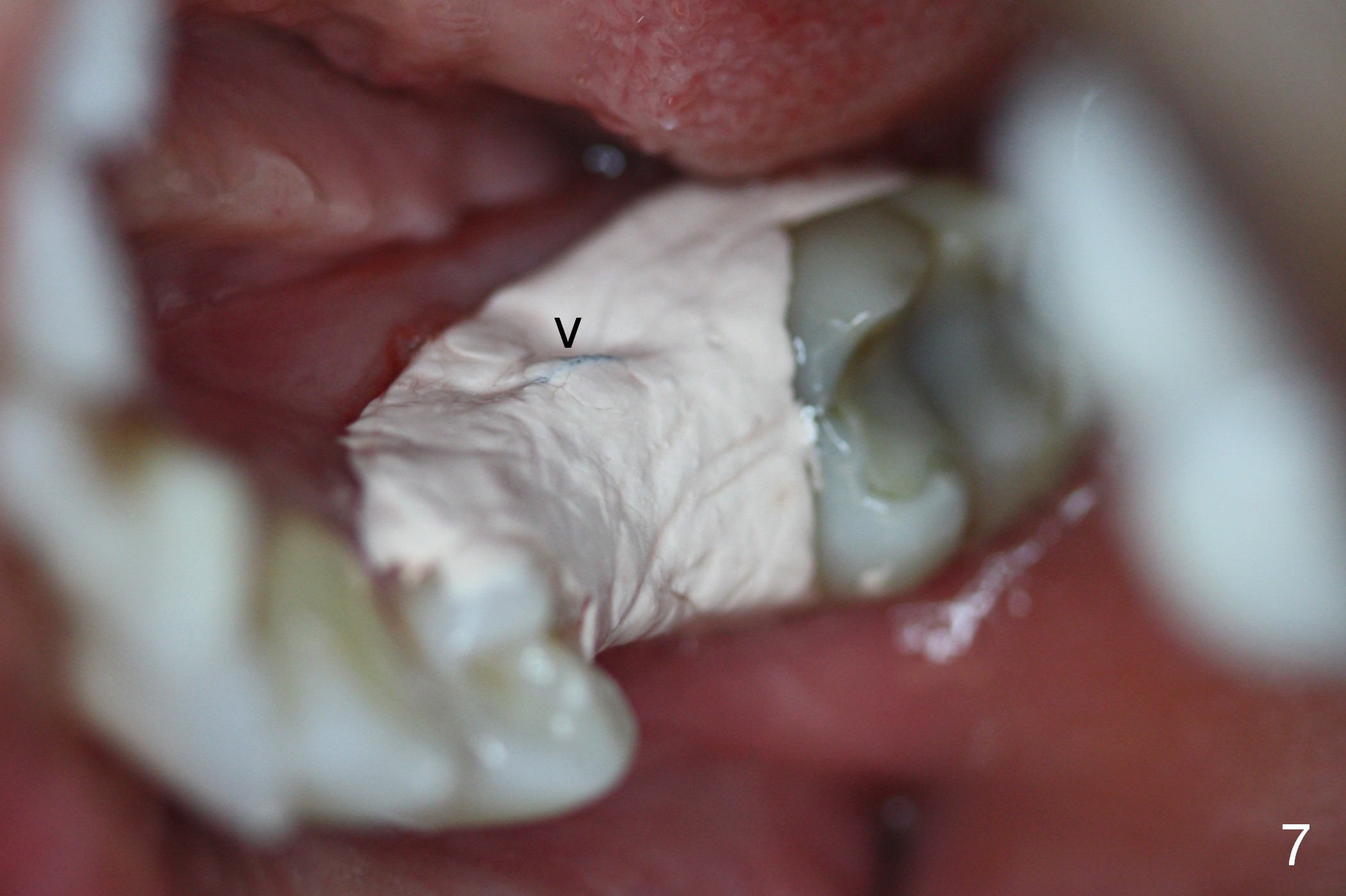
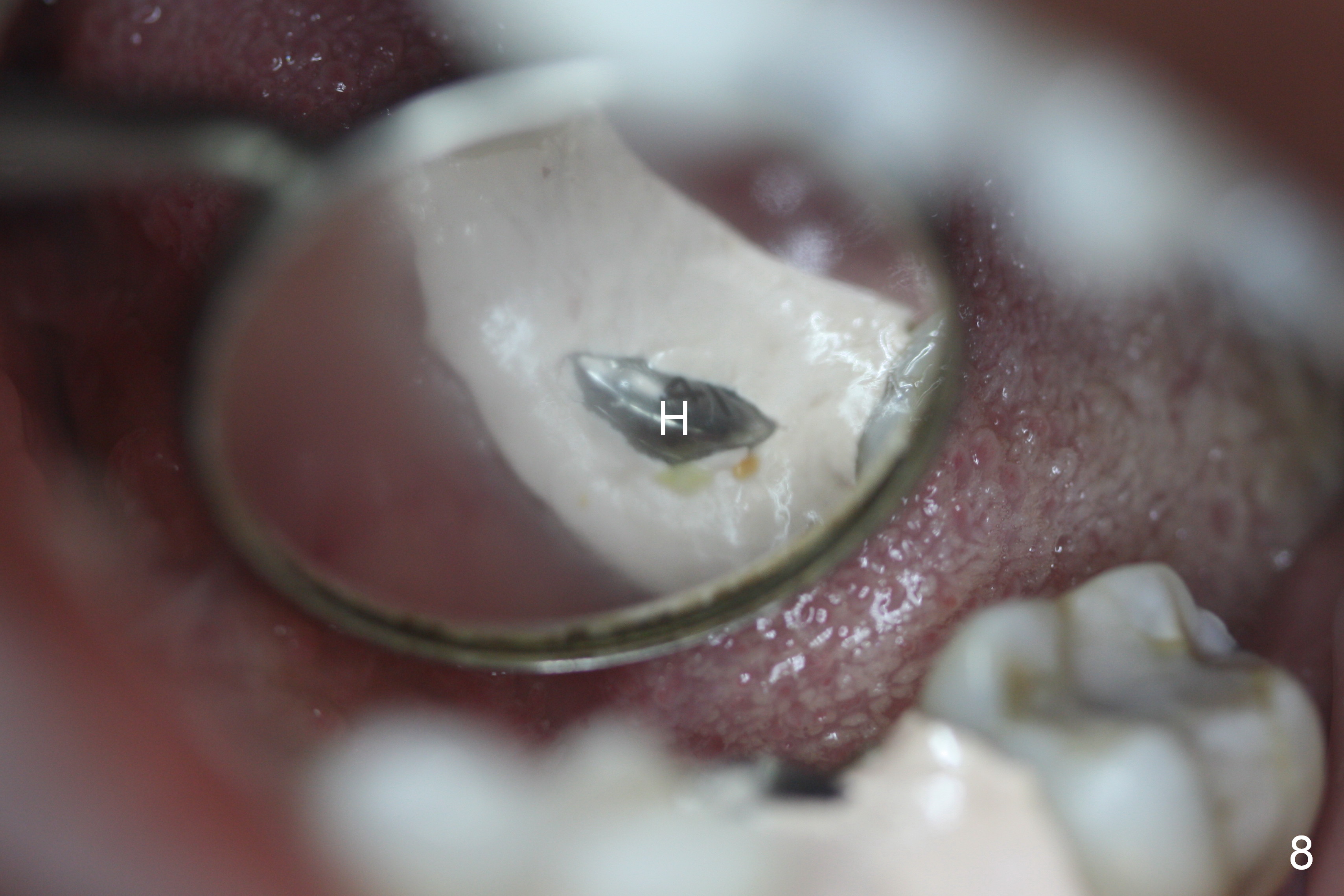
Hassel Associated with Delayed Implant
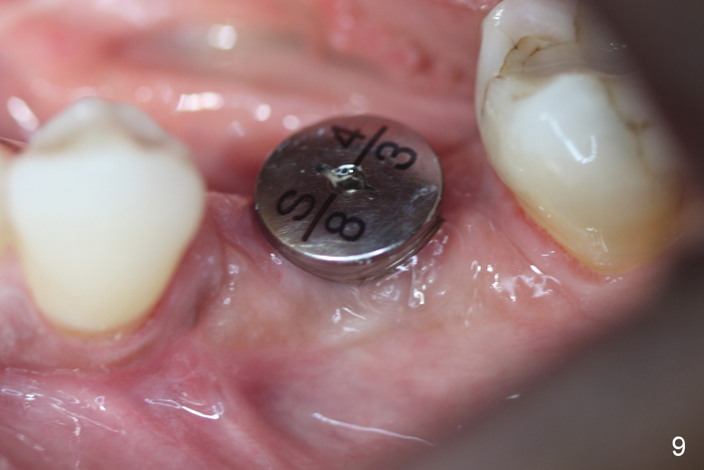
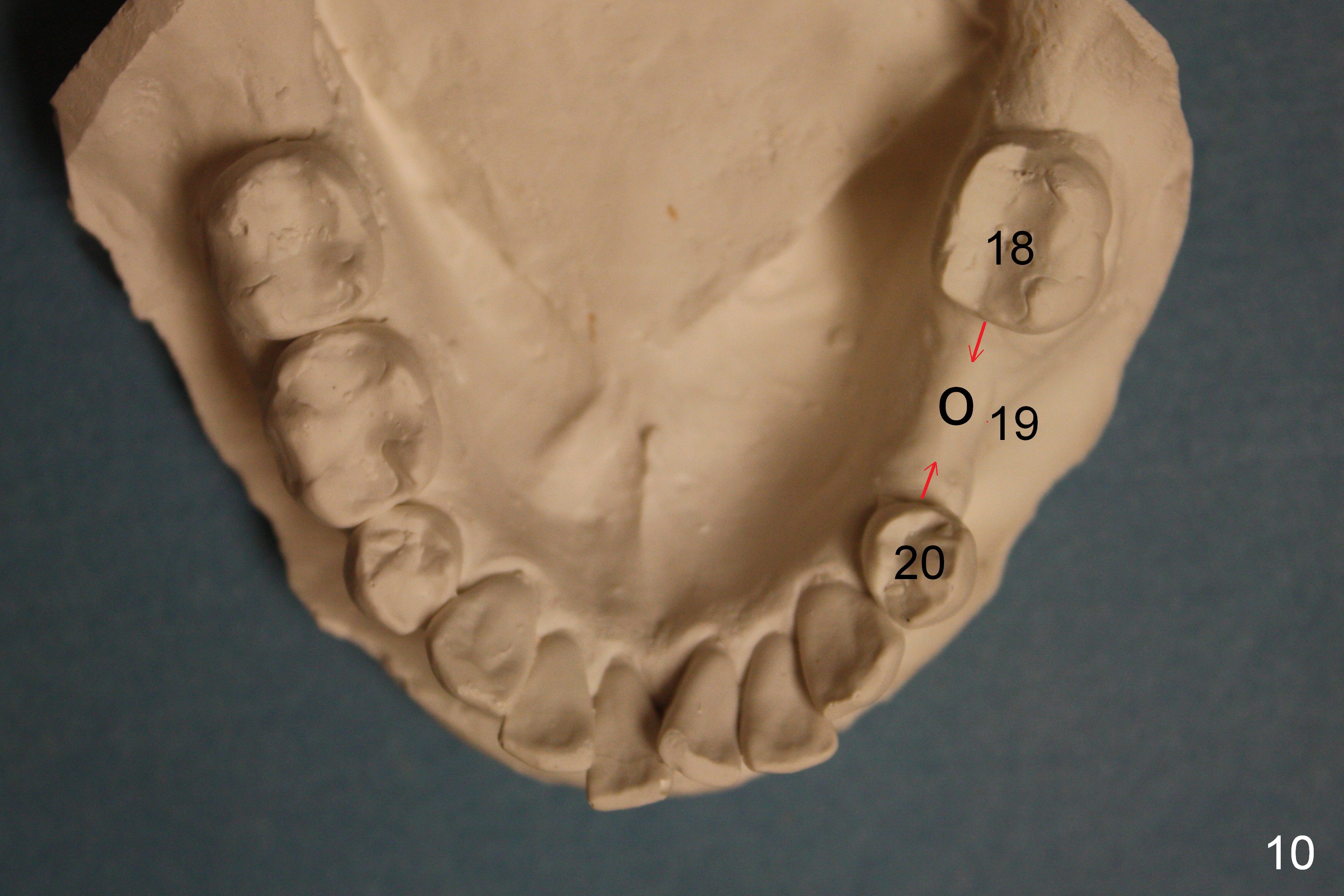
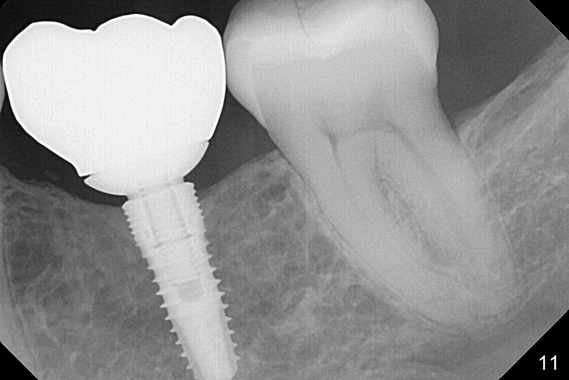
A 44-year-old black lady is a typical dental phobic. She has lost #19 for years with history of traumatic extraction. The ridge is atrophic (Fig.1). She is not comfortable with intraoral X-ray. So CBCT is taken. The coronal section shows that the bone density is low in the coronal ridge (Fig.2 arrowheads). It is difficult to determine the buccolingual width at crest; probably a 5.9x10 mm implant is appropriate (Fig.3). The most useful information from CT is height determination for the implant (Fig.4). The initial osteotomy depth is 10 mm (Fig.5); there is a large safety margin. There is a 2 mm clearance when a 4.5x12 mm implant is placed (Fig.6). Incision is sutured; a 8x4 (3) mm healing abutment is placed (Fig.7 arrowhead), followed by application of perio dressing. The latter remains stable 13 days postop thanks to the holding effect of the healing abutment (Fig.8 H). The wound has apparently healed when the dressing is removed (Fig.9). It appears that the edentulous space is extremely wide. The 4.5 mm implant may not sustain masticatory force. Limited orthodontic treatment appears to be necessary, using the implant as an anchorage to move the neighboring teeth (Fig.10 arrows). In fact the orthodontic treatment is not rendered. A crown is cemented 14 months postop. The cortical bone is thickened around the implant 15 months post cementation (Fig.11).
Return to Professionals,
Lower Molar Immediate Implant,
Dr.
Wu
Xin Wei, DDS, PhD, MS 1st edition 04/15/2015, last revision 09/02/2017