|
|
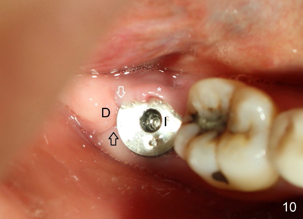
Fig.10: The abutment is removed to reveal the implant (I) and excess lingual gingiva (*). The buccal (black arrow), lingual (white arrow) and distal (D) gingivae have healed each other, as compared to Fig.7.
Return to No Drill Implantation
Xin Wei, DDS, PhD, MS 1st edition 10/10/2014, last revision 09/13/2015