 |
 |
 |
 |
 |
Implant Placed in the Mesial Socket of the Lower 2nd Molar
Sometimes the lower 2nd molar has two roots. An implant may be placed in the mesial socket. The occlusal table of the future crown can be fabricated narrow mesiodistally to reduce lateral force. It may be easy for oral hygiene to be done distally.
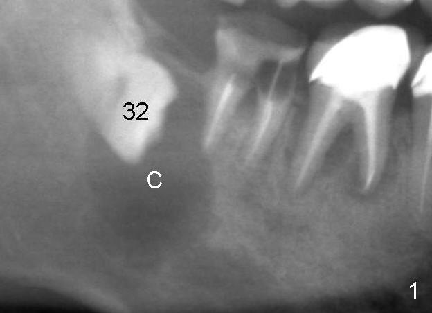
A 47-year-old man has poor dentition (Fig.1). The tooth #31 is non salvageable. It has two roots. There is a dentigerous cyst (C) associated with #32. There is no apparent bone distal to the distal root of the 2nd molar.
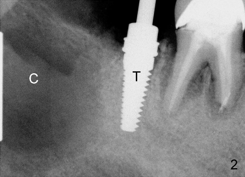
Immediately post extraction and cyst enucleation, osteotomy is formed in the mesial socket of the 2nd molar surrounded by intact bone (Fig.2 (T: 5x17 mm tap)). Separation of the inferior alveolar neurovascular bundle from the cyst causes severe hemorrhage, which is stopped temporarily by gauze.
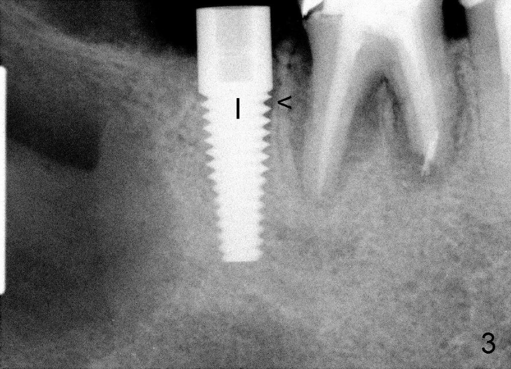
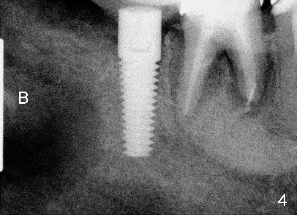
A 5x17 mm implant is placed (Fig.3). When the gauze is removed, copious hemorrhage resumes, which is stopped by pressing into the defect with mixture of autogenous bone (harvested from osteotomy in the mesial socket of the 2nd molar) and 3.5 g of allograft (Fig.4: B).
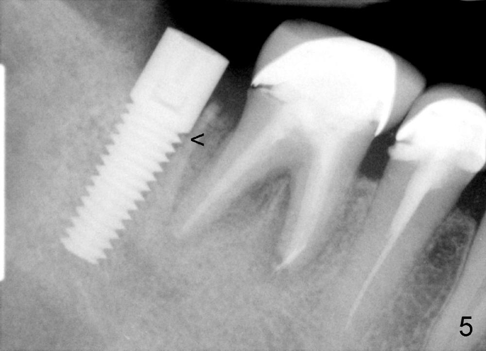
Three and a half months postop, the density of the previous cyst area increases apparently, while the bone grows into the threads of the implant (Fig.5 <, as compared to the same area of Fig.3,4). The implant is ready for restoraton.
Xin Wei, DDS, PhD, MS 1st edition 01/29/2014, last revision 01/19/2018