.jpg)
|
|
|
|
|
|
|
|
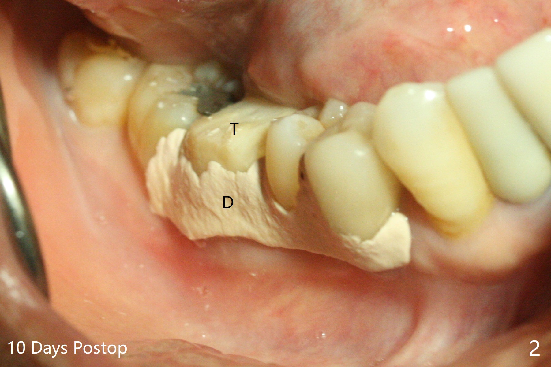
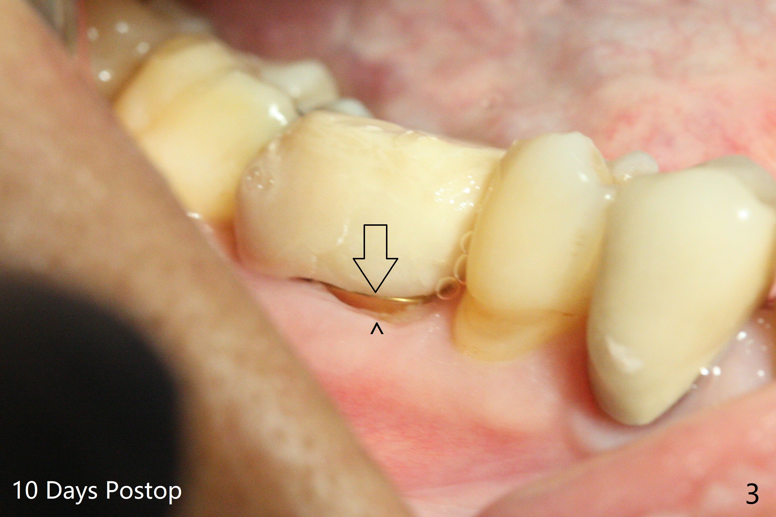
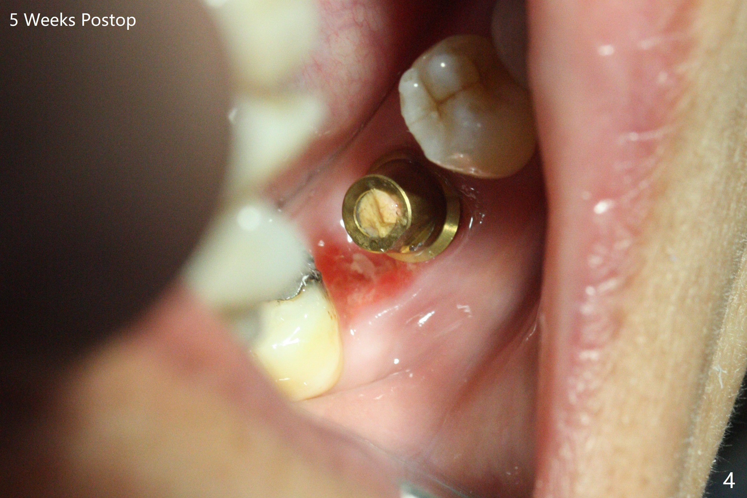
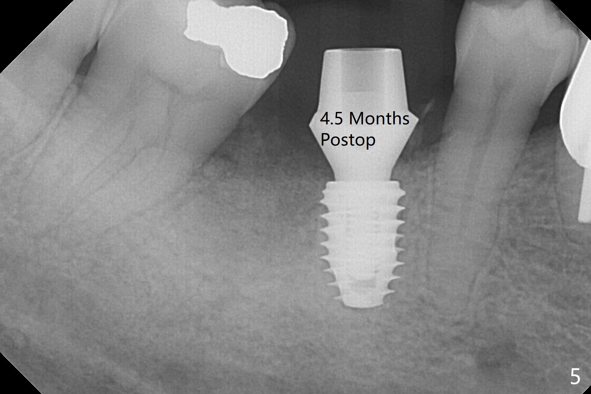
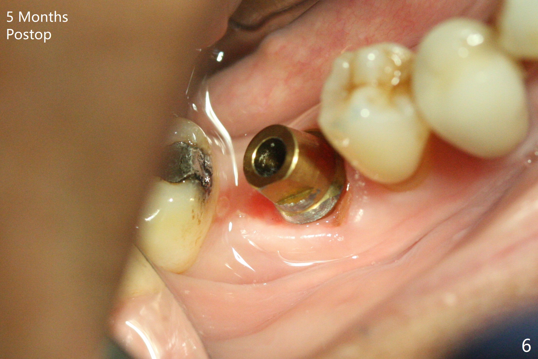
Stable and Precise
In spite of severe bone loss and 1.86 mm remaining bone after extraction of the tooth #30, a 5x7.3 mm implant achieves insertion torque of 35 Ncm with an immediate provisional (Fig.1). Periodontal dressing is applied for additional fixation of the bone graft (*) and Osteogen plug (P). Although the implant is placed in the mesial socket, the coronal end of the abutment is in the middle of the edentulous area. The patient is extremely pleased with no pain surgery. There is no postop paresthesia. The periodontal dressing (Fig.2 D) remains in place and buccal to the temporary crown (T) 10 days postop. When the former is removed, there is a gap between the margin of the provisional and that of the gingiva (Fig.3 between arrow and arrowhead), suggesting that the latter has shrunk postop. If there were no periodontal dressing, some of bone graft may have been lost. The distal socket heals when the immediate provisional is removed for revision 5 weeks postop (Fig.4). With gingival retraction cords, the abutment margin is prepared 2 months postop, particularly distal, to reduce food impaction in the future. If there is no abutment screw loosening with final restoration, it suggests that computer designed trajectory is acceptable. The implant plateau seems to be covered by the bone 4.5 months postop (Fig.5). In fact the mesiobuccal margin of the abutment is close to the corresponding crestal bone. The gingiva looks healthy 5 months postop (immediately before cementation, Fig.6), although MO amalgam of the tooth #31 is breaking down (*). Return to Lower Molar Immediate Implant, Prevent Molar Periimplantitis (Protocols, Table), Trajectory Xin Wei, DDS, PhD, MS 1st edition 04/17/2019, last revision 09/14/2019