.jpg)
%20at%2029.jpg)
|
|
|
|
|
|
|
|
|
|
|
|
|
|
|
|
|
|
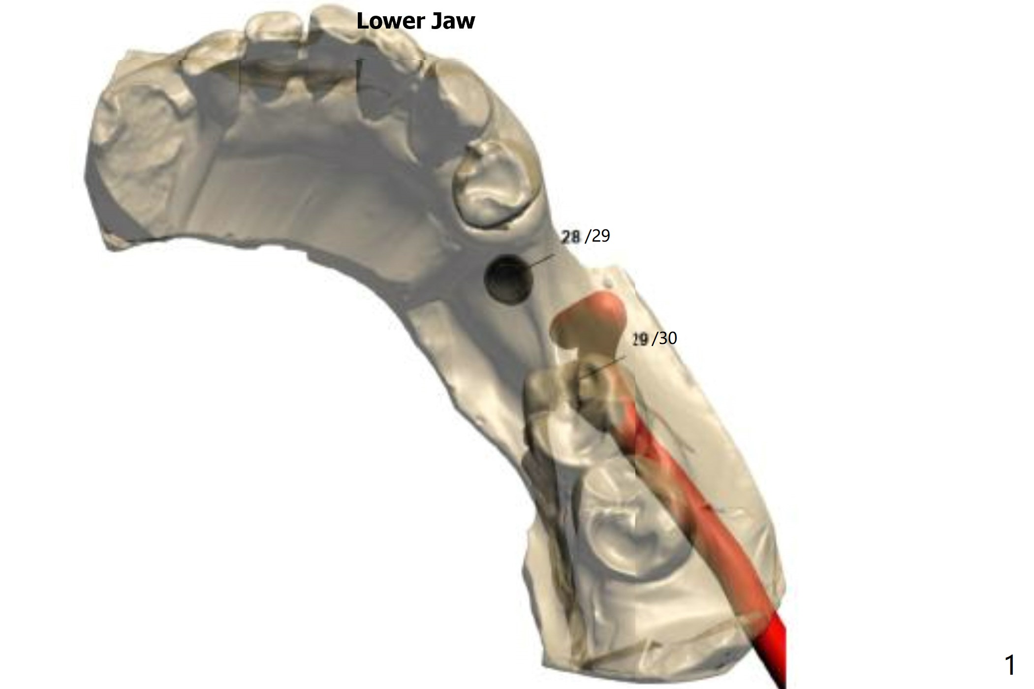
Nonkeratinized Gingiva
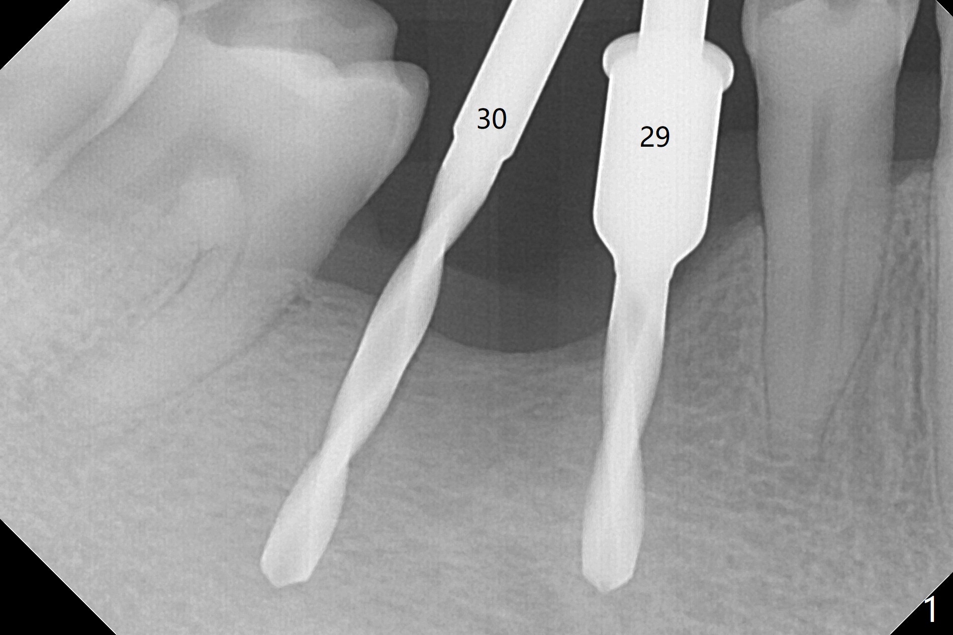
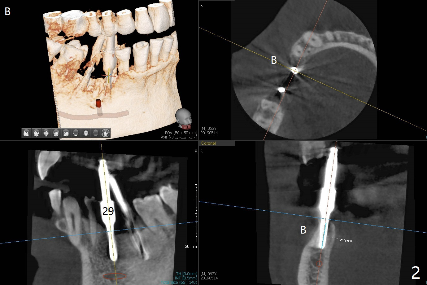
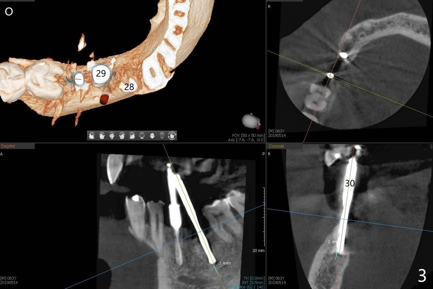
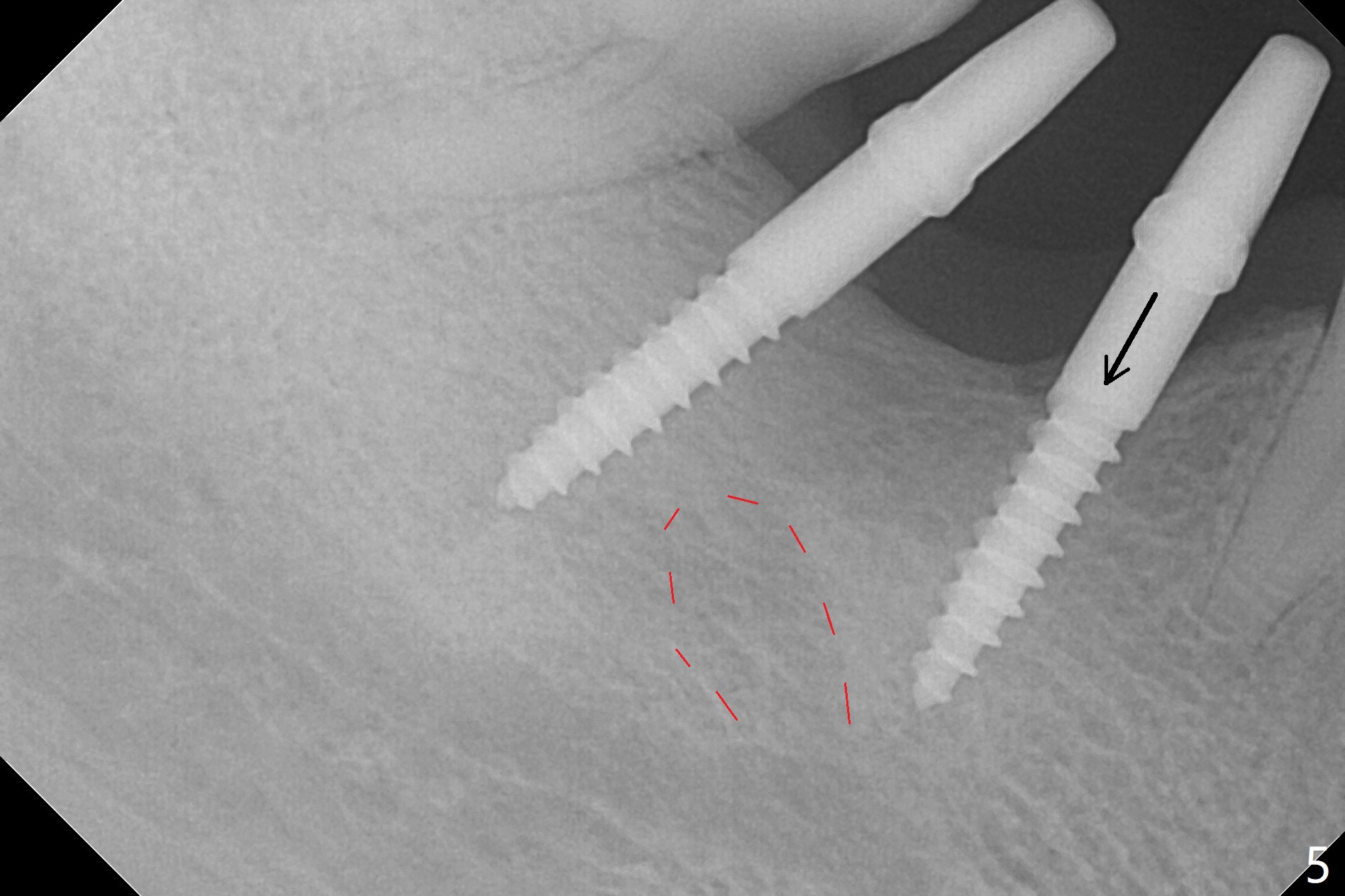
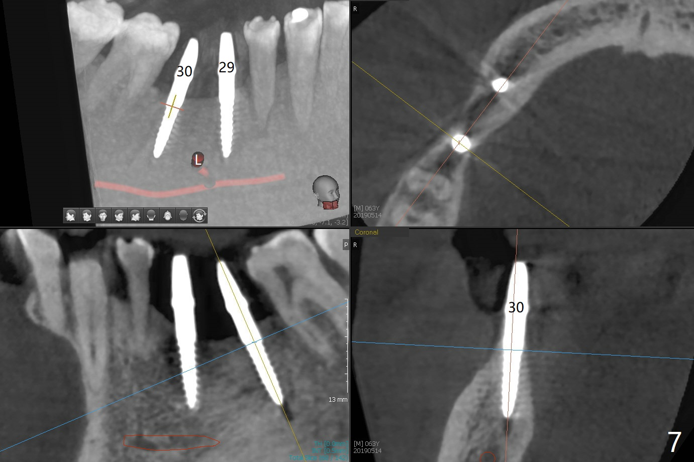
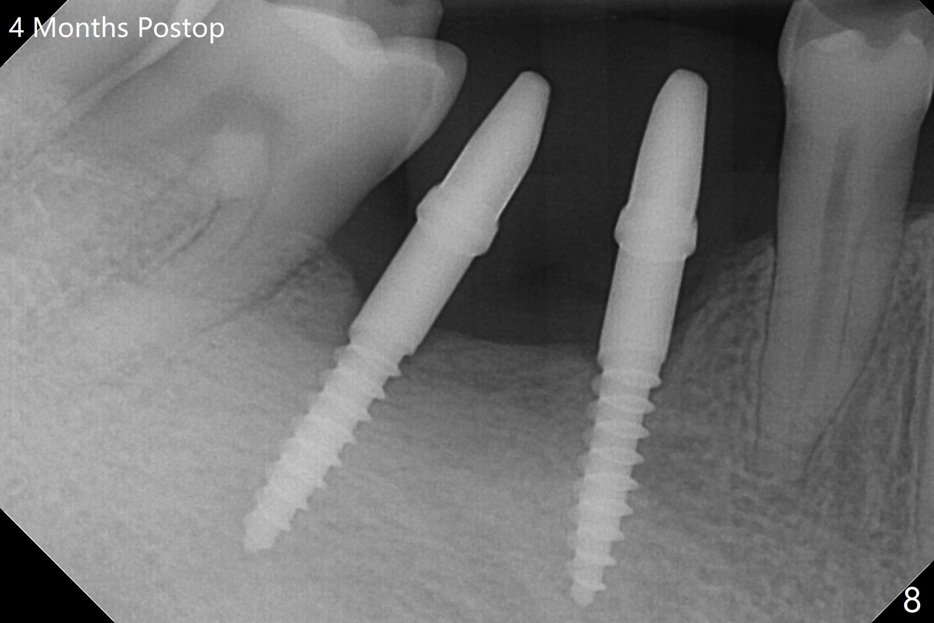
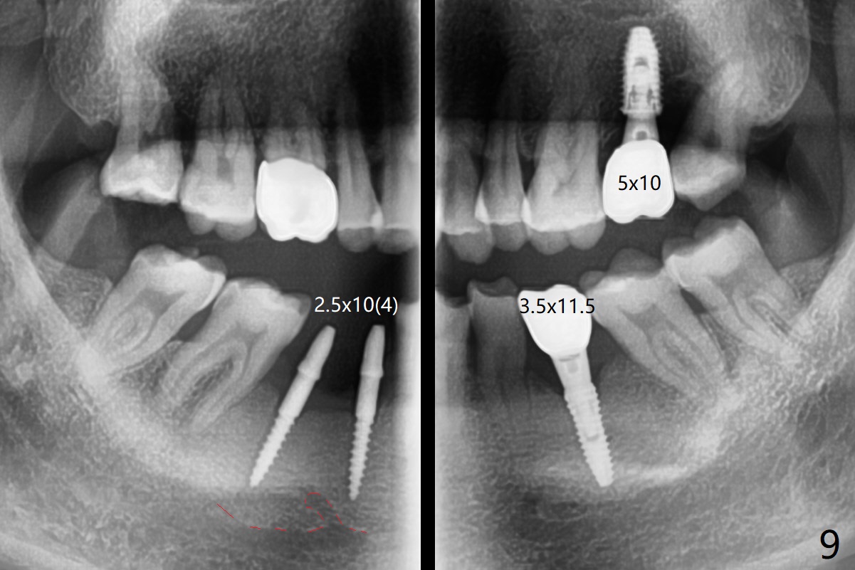
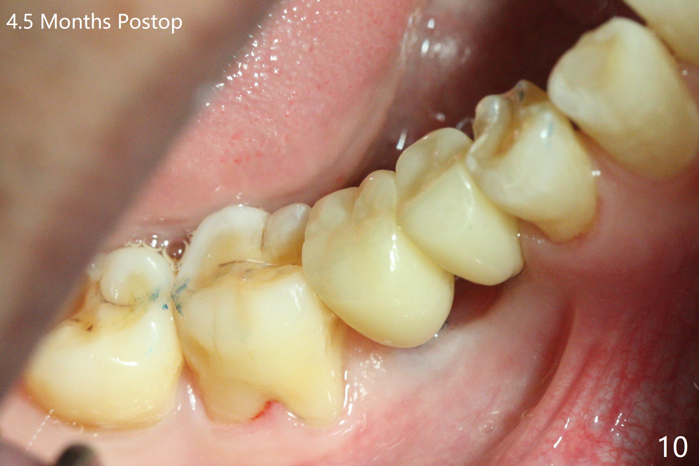
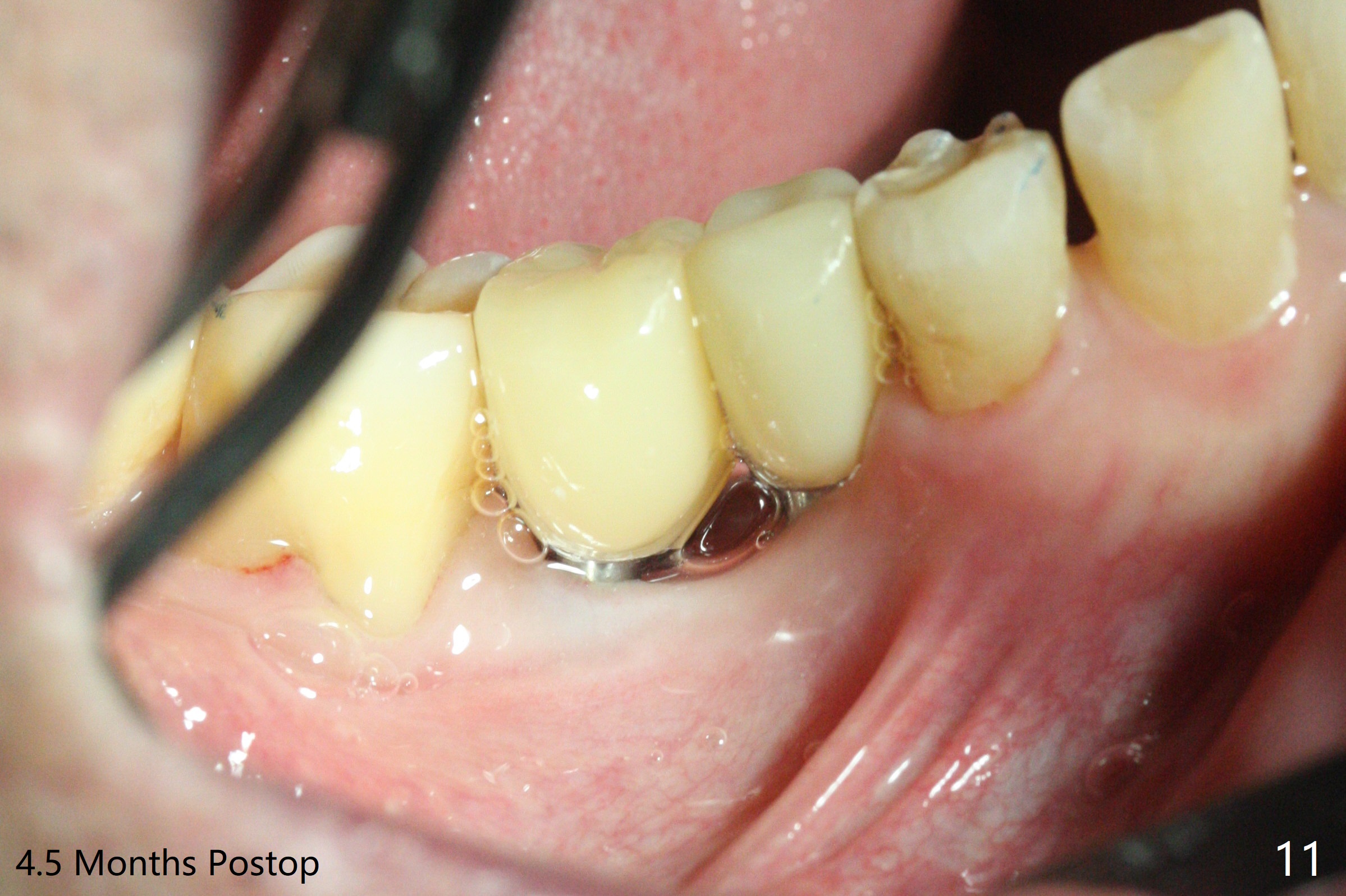
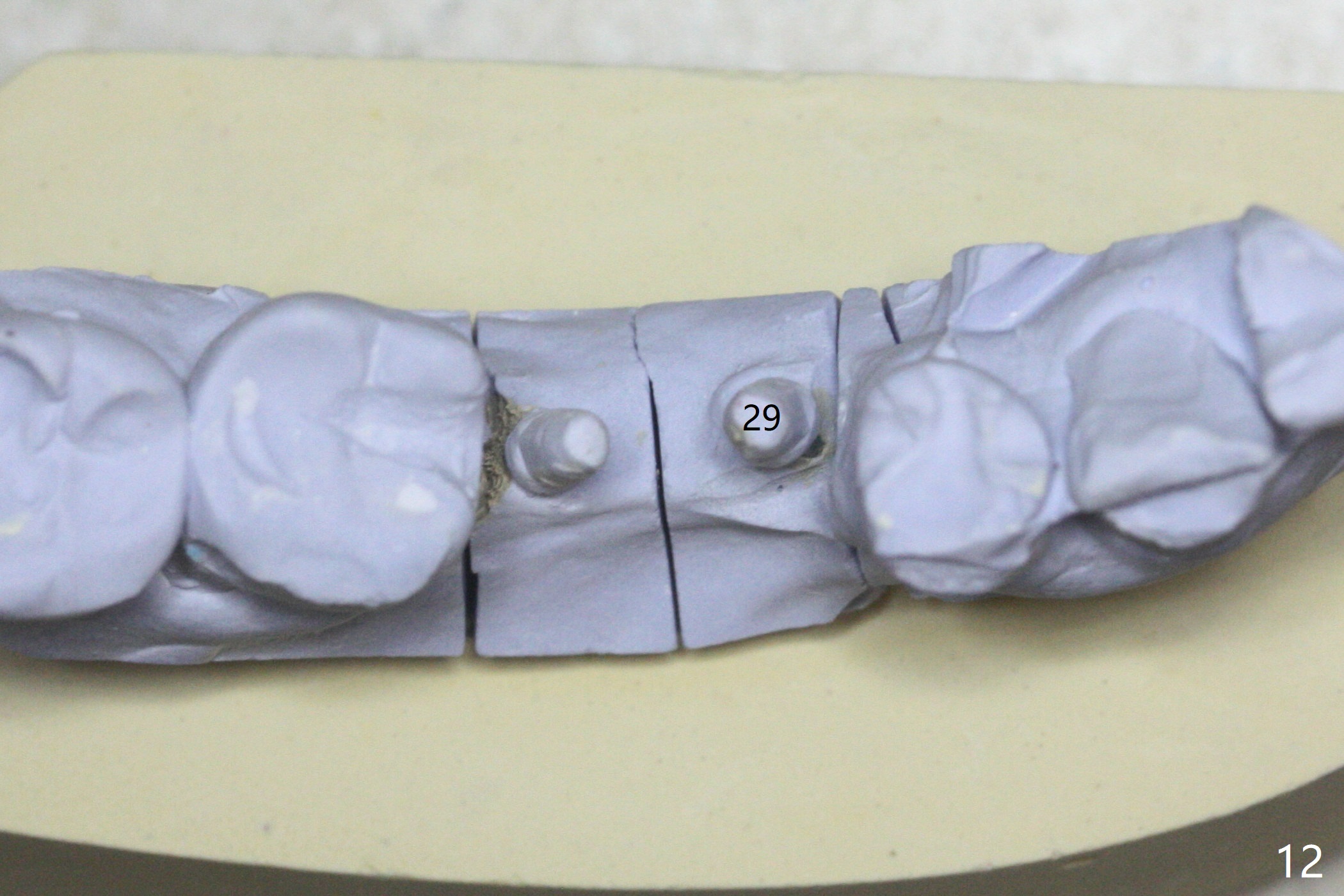
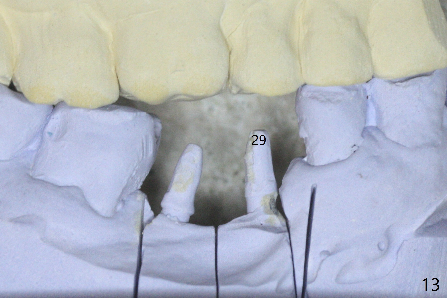
After initial osteotomy using 2.2 mm drill for 10 and 11.5 mm at #29 and 30, respectively, the guide is removed for re-insertion of drills and PA (Fig.1). The osteotomy at #29 is found lingually and in the nonkeratinized gingiva with mild laceration (wish incision to be made before osteotomy); the osteotomy is established a little lingual at #29 (Fig.2) and more or less in the middle of the narrow ridge and #30 (Fig.3). Lindamann bur is used to move #29 osteotomy buccally. A 1.5 mm drill is used to increase the depth free hand before placement of 2.5x10(4) mm 1-piece implants (Fig.4). After adjustment of the implant depth (Fig.5), CT is retaken, which shows proper implant placement (Fig.6,7). Probably due to good oral hygiene, the gingiva around the implants appears to be keratinized 4 months postop (Fig.8). After abutment preparation for margin and parallelism, impression is taken (Fig.9). When the permanent crowns are temporarily cemented, the large gingival embrasure is noted (Fig.10,11). The latter could be reduced by modifying the provisional in the healing stage. In fact the crown at #29 dislodges 2 days post cementation because lingual (Fig.12) and distal (Fig.13) placement. A surgical stent should have been fabricated from RPD for free hand placement!
Return to
Lower Molar
Immediate Implant,
Trajectory
Xin Wei, DDS, PhD, MS 1st edition
05/14/2019, last revision
10/05/2019