|
|
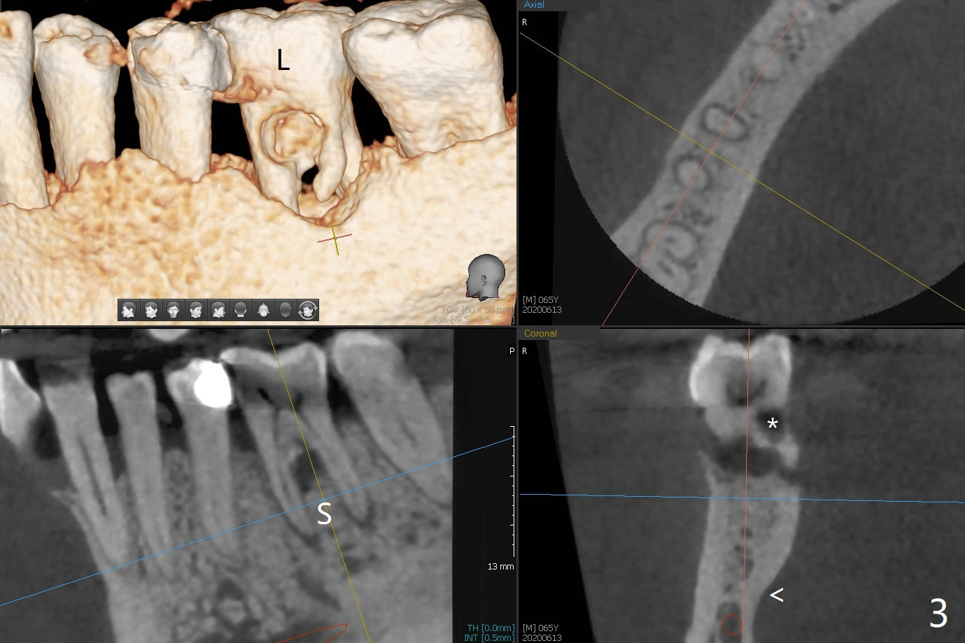
CT shows the submandibular fossa (Fig.3 <), dictating a short implant 10 mm, Fig.4). To place the implant in the septum (Fig.3 S), the coronal portion of the tooth is removed (Fig.5 black area) so that the roots are able to keep the osteotomy without deviation (Fig.6 red arrow). *: caries; L: lingual.
Xin Wei, DDS, PhD, MS 1st edition 06/13/2020, last revision 06/13/2020