|
|
|
|
|
|
|
|
|
|
|
|
|
|
|
3rd Molar Kept for Guide Distal Anchor
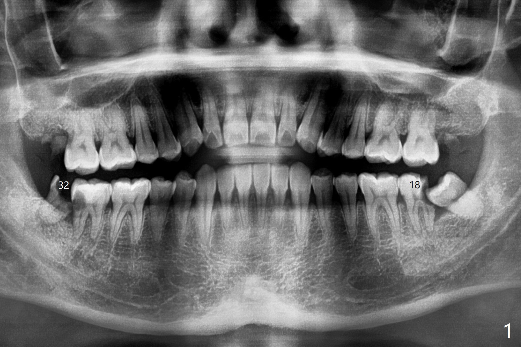
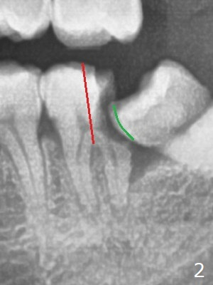
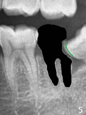
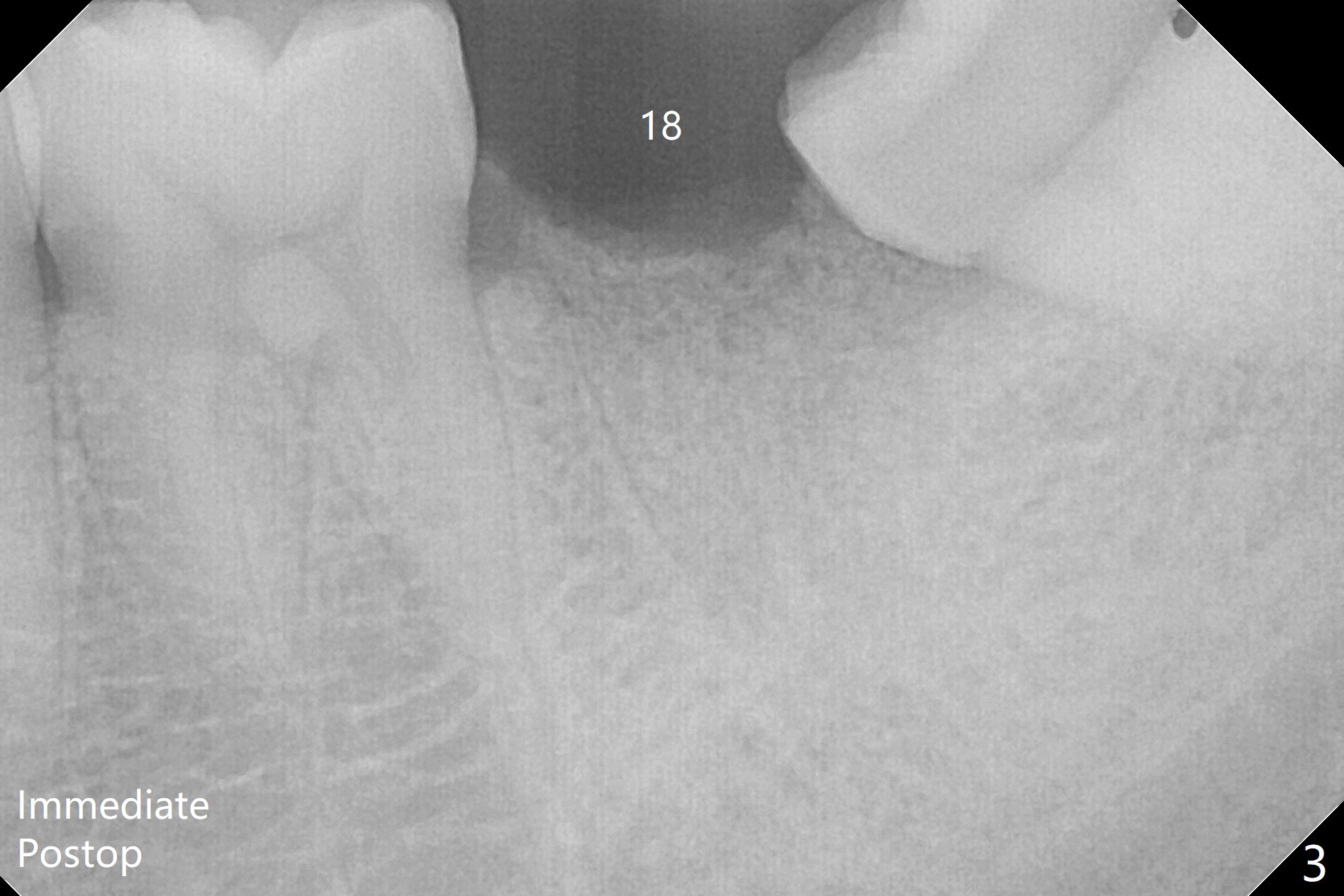
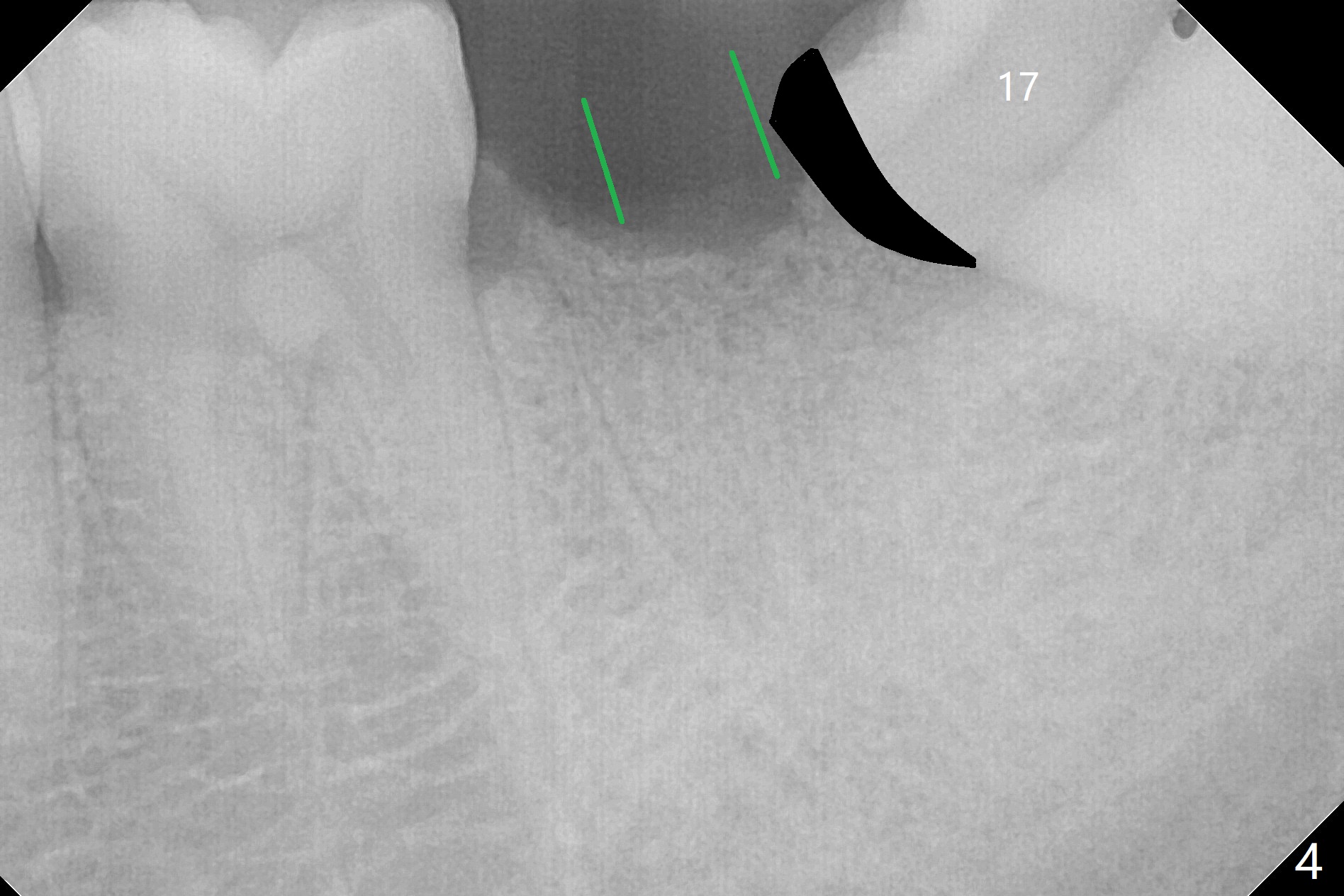
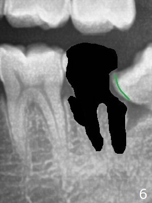
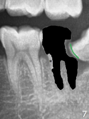
A 36-year-old man with poor dentition (smoker 1/4 ppd) requests extraction of sympto-matic teeth (#18 and 32, Fig.1). A half of Osteogen Plug is placed at #32 post extraction. To extract #18, the tooth has to be sectioned (Fig.2 red). The mesial portion is removed easy, while the distal one requires removing the mesial surface of #17 (Fig.2 green). Vanilla bone is placed after extraction (Fig.3), covered by 8x8 mm Amnion-Chorion Allograft and sutured with 4-0 PGA. Four months later, the mesial surface of #17 will be trimmed (Fig.4 black area) so that surgical guide metal sleeve (green) will be seated in the neutral position of the edentulous area. After extraction (Fig.5 black), the distal portion of the mesial crest will be resorbed, leading to gingival papilla atrophy and food impaction (Fig.6). Keeping the mesial portion of the root (Fig.7 *, socket shield) is able to prevent mesial crestal bone loss. The immediate implant will be placed ~ 2 mm short of the depth and checked whether it touches the retained root or not.
Return to
No Deviation
Plug
Socket Shield
Xin Wei, DDS, PhD, MS 1st edition
03/06/2020, last revision
04/27/2020