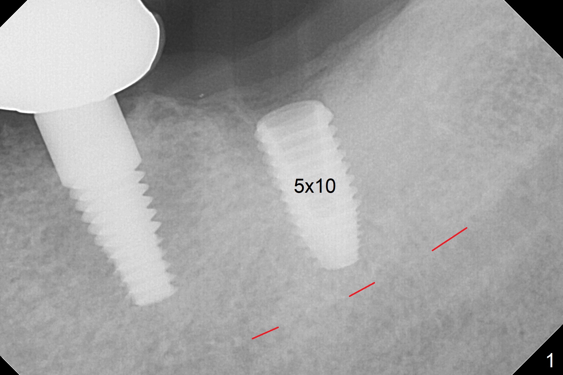
Depth Control of Guide for 2nd Molar
When the surgical guide is pressed hard at the 1st molar area, the guide
appears to sink down ~ 1 mm. The last drill and a 5x10 mm implant are
placed as the guide is being pressed hard (Fig.1). The implant appears
placed deeper than the
design.
Hello Dr. Wei,
Thank you for your feedback.
I tried to merge the original planning to the panoramic view you sent me.
However it did not work out as the angle you took the pano is quite leaning
toward buccal, not perpendicular. So I could not merge those to see the
difference.
So, here's my question, what is the distance you got from the distal of #19
to the #18 implant? I think the distance is different from the original
planning too.
In order to identify which point to improve for the next cases, it would be
helpful for us to have that information.
And for the free-end case like this, we recommend drs to hold the guide
firmly where the remaining teeth are located. But not too hard but with
enough strength to make the surgical guide stable not rocking. When
necessary, you may need to press 2 points on the surgical guide with 2
fingers and holding the patient's chin. And for free end case, most dentists
tend to have the implant contra angle towards operator which means implant
apex will be distalized and the top part of implant will be mesialized as it
is hard to put long drills into distal area such as molars. But most
importantly, the path contra angle should be perpendicular to the metal
sleeve so that it could follow the originally planned path.
If there's anything I am missing please let me know so that I could get the
better answer for you.
June
20, 2018 7:54 PM
Thanks, Jennifer. I appreciate the method holding the guide with 2
fingers instead of one and also holding the chin, which I missed. X-ray I
presented to you is PA. It does tilt. The way I pressed the guide
somewhat changed the osteotomy distal. Thanks again for the input.
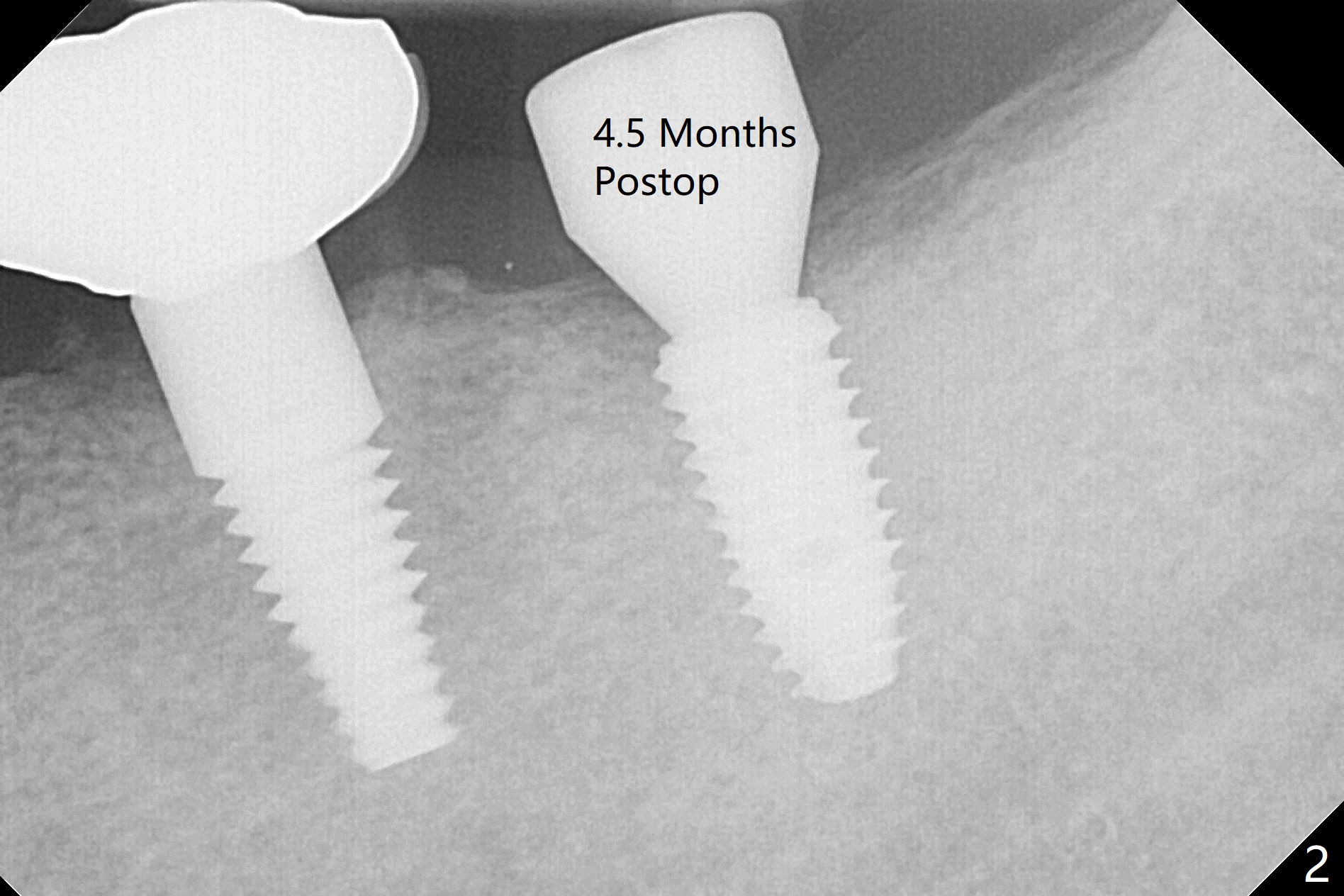
Crestal bone density increases 4.5 months postop (Fig.2).
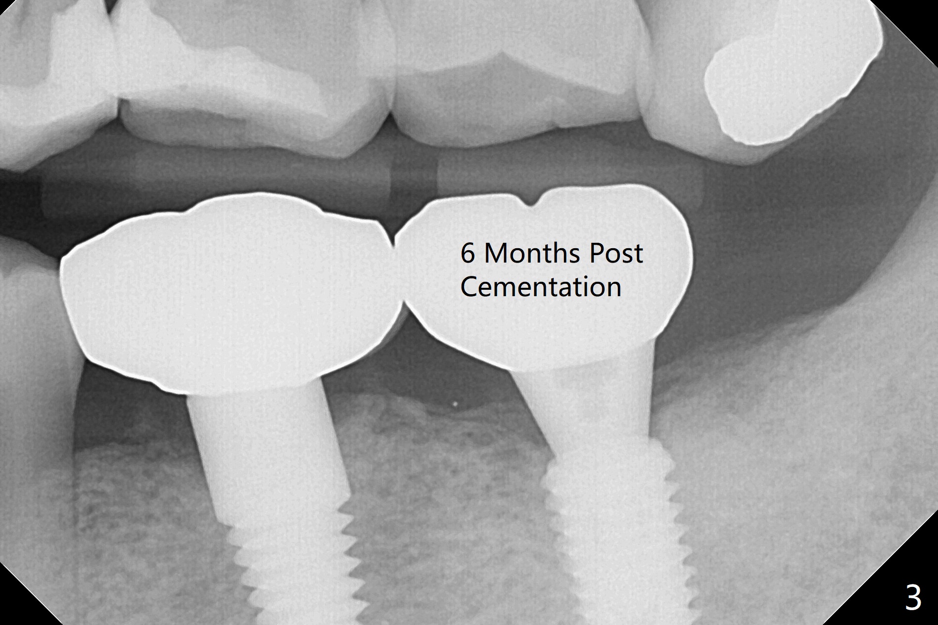
There is no bone loss 6 months post cementation (Fig.3 (BW)).
Return to
Lower
Molar Immediate Implant,
Armaments Xin Wei, DDS, PhD, MS 1st edition 06/19/2018, last revision
06/02/2019