|
|
|
|
|
|
|
|
|
|
Socket
Preservation for Lesion Extending to IAC
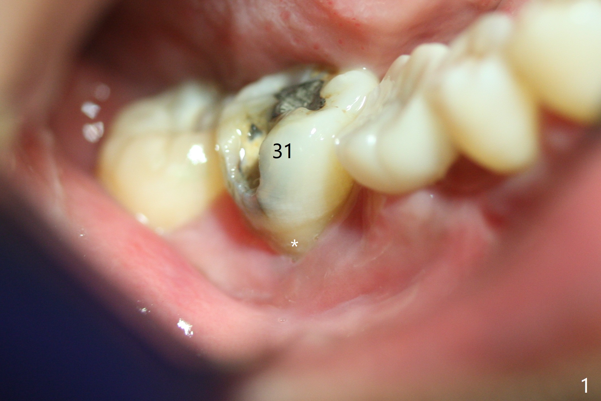
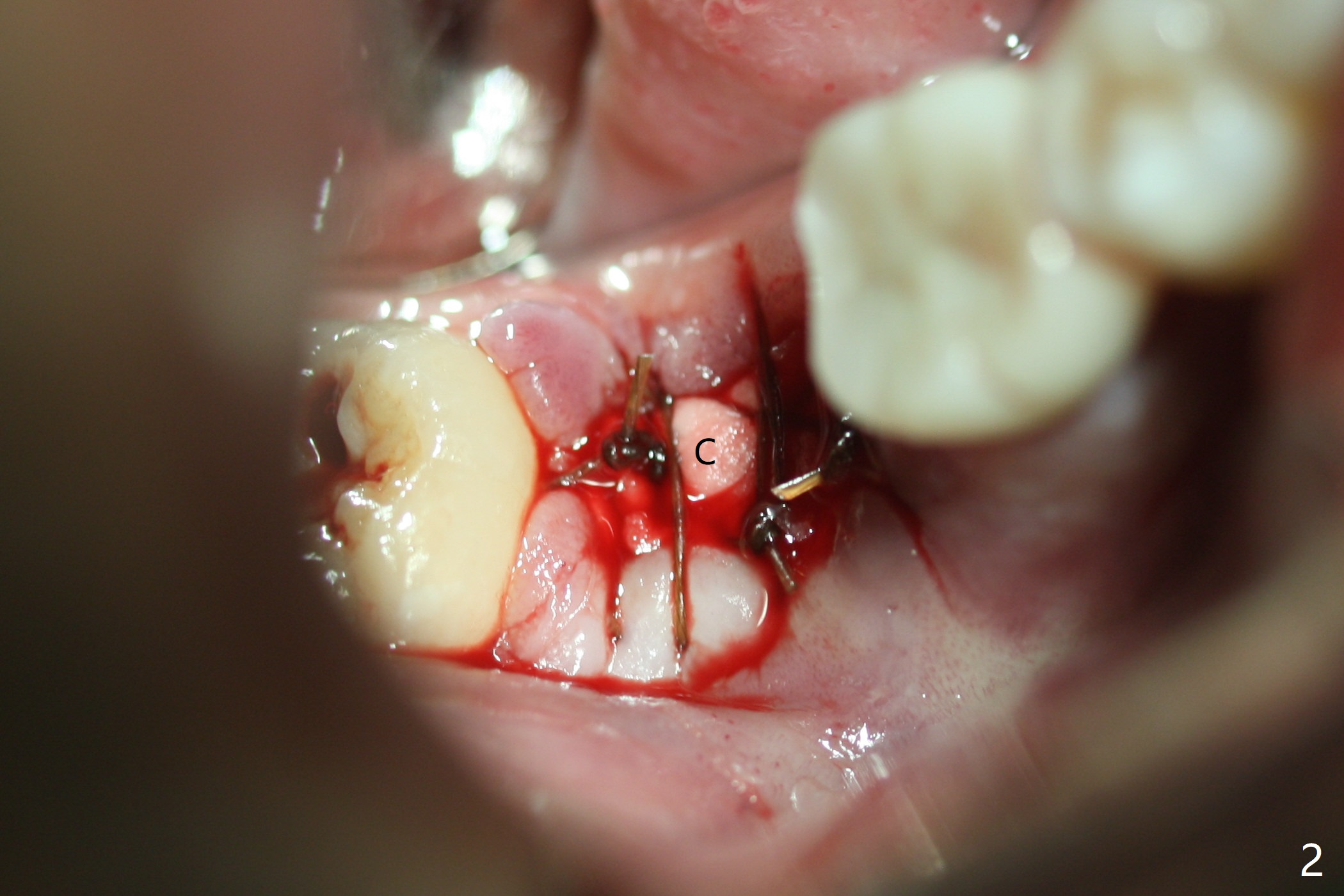
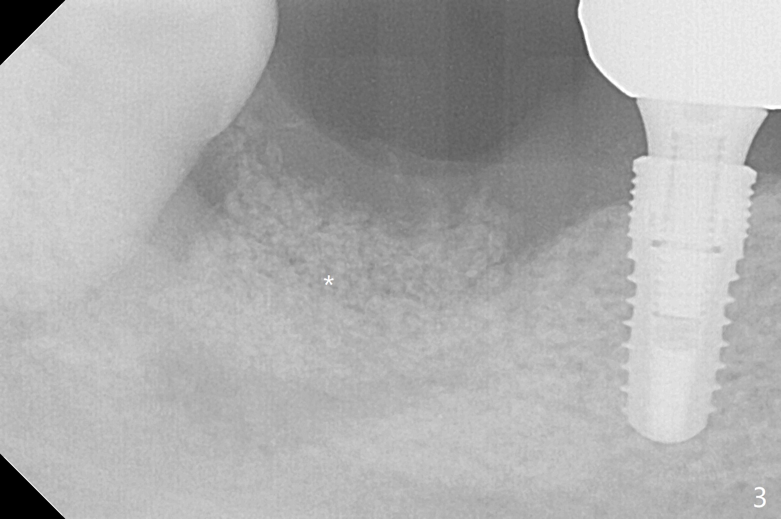
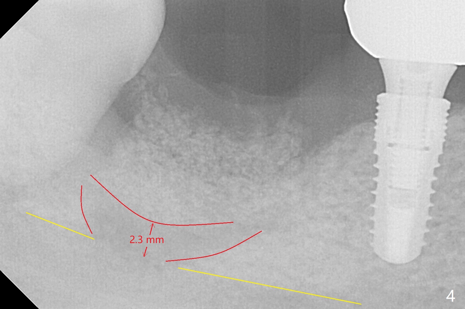
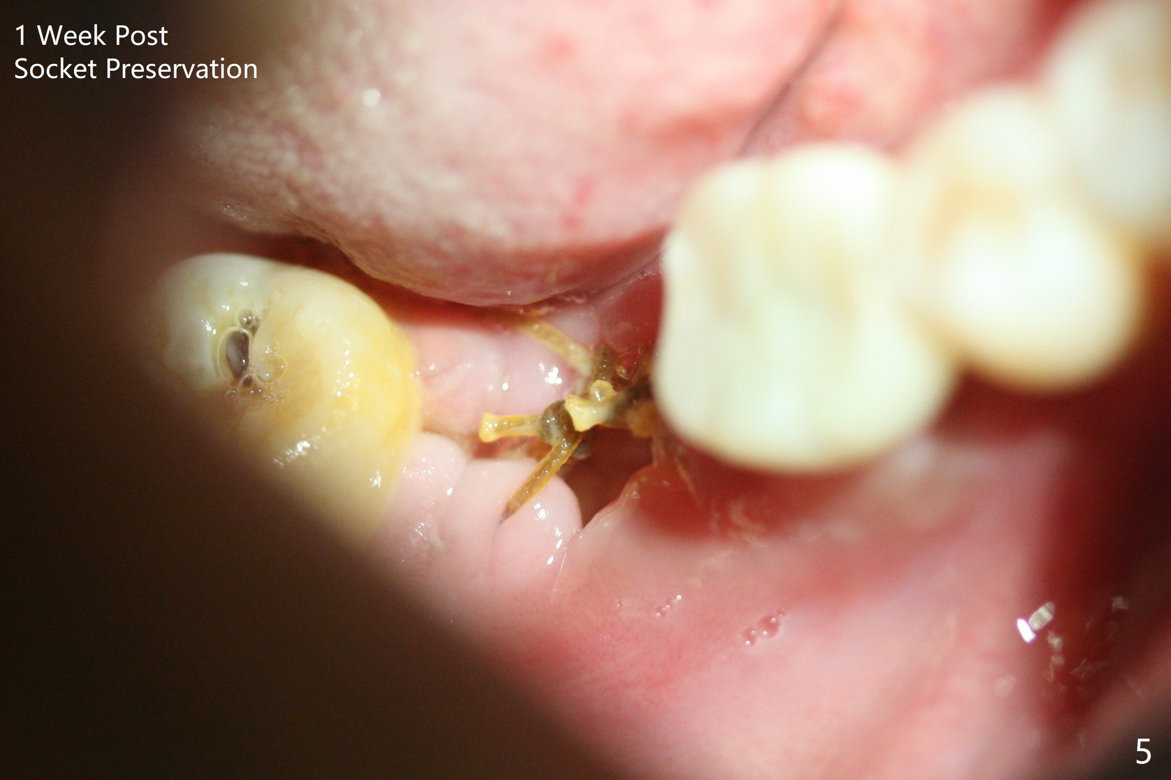
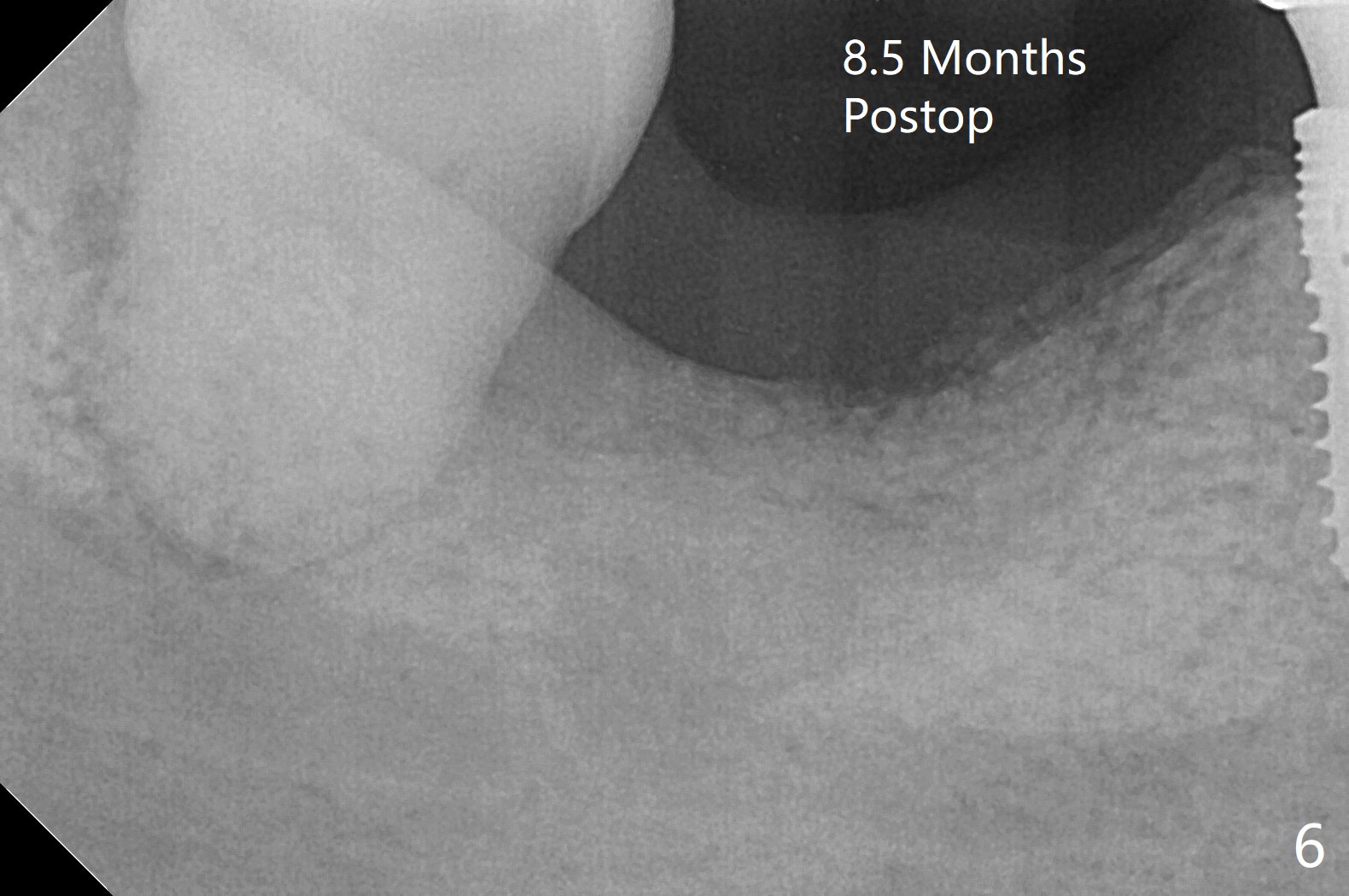
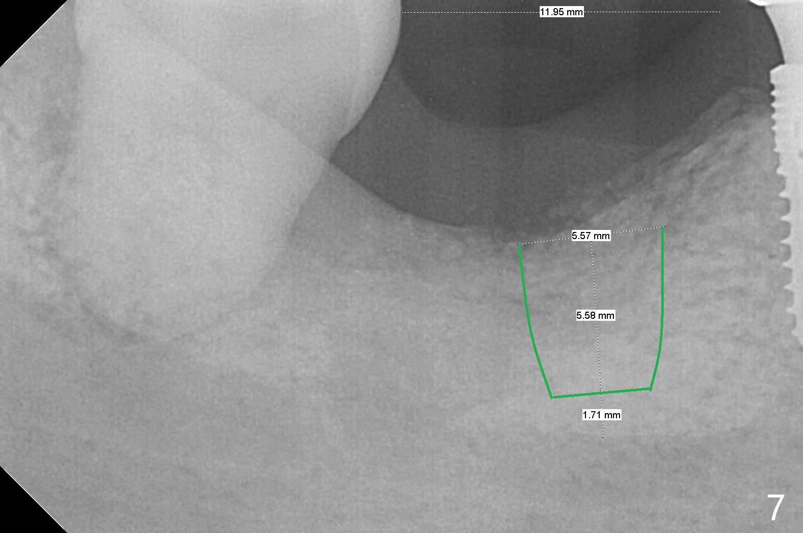
Although the bony socket is shallow, the soft tissue one remains deep at #31 with gingival recession (Fig.1 *). The deep soft tissue socket with hemorrhage after extraction presents difficulty in debridement of granulation tissue apically. After placement of allograft (Fig.3 *) and overlying collagen membrane (Fig.2 C), the socket is closed with 4-0 Chromic gut suture. In fact, there is ~ 2 mm granulation tissue left (Fig.4 red (yellow line: upper border of the Inferior Alveolar Canal)). To reduce socket shrinkage, the tooth #32 is not extracted. The collagen membrane has lost 1 week postop (Fig.5). More sutures should have been used, preferably using Human Amnion Chorion Allgraft as a membrane. The latter promotes wound healing. The bone graft also seems to have lost in 8.5 months (Fig.6). A short implant will be placed mesially and obliquely (Fig.7).
Return to
Lower Molar
Immediate Implant,
Trajectory,
Wechat
Xin Wei, DDS, PhD, MS 1st edition
01/08/2019, last revision
09/24/2019