|
|
|
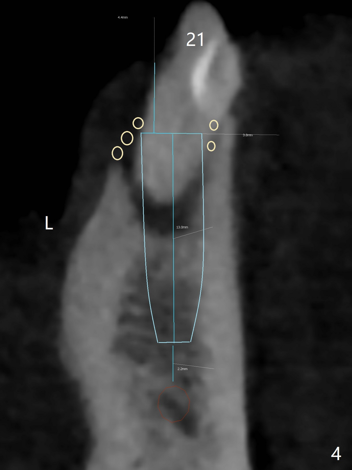
The Inferior Alveolar Canal (Fig.5 red circle) becomes the Incisive Canal (IC, Fig.4 red circle) after the Mental Loop (Foramen). Damage to IC has less consequence (without neurologic deficit), but it may cause severe intraoperative hemorrhage. There should be sufficient clearance during osteotomy (Fig.4 ~ 2mm).
An implant at #21 (3.5 or 4x13 mm) will be placed at the level between the buccal and lingual crests (Fig.4) with osteotomy initiated in the middle of the socket. Bone graft is to be placed around the coronal end of the implant (yellow circles).
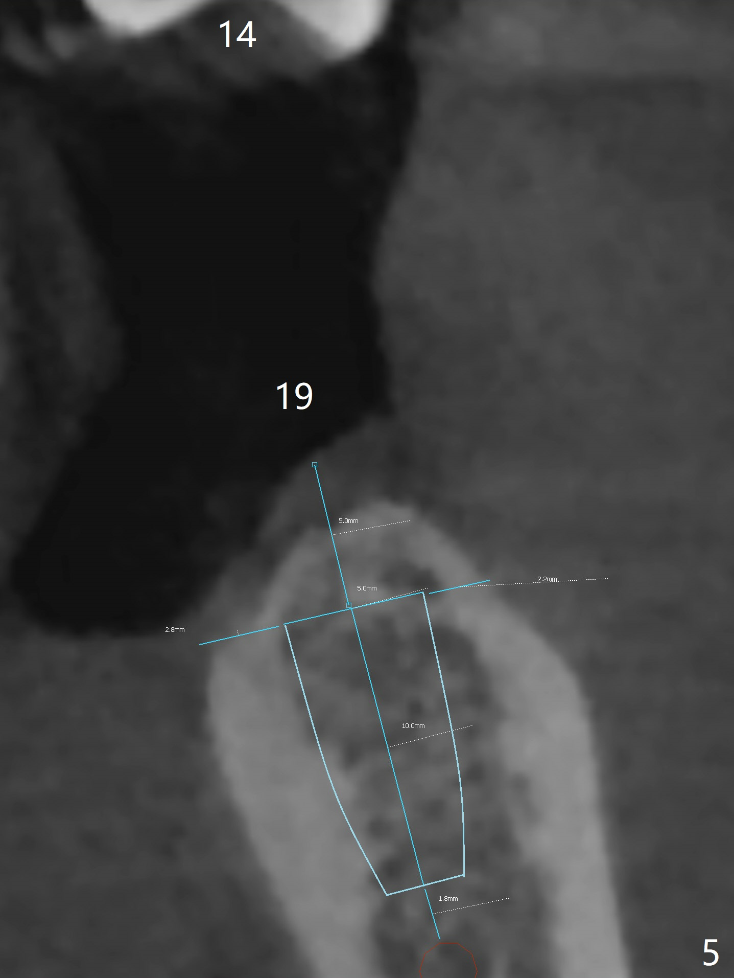
The socket at #19 appears to have not completely ossified (Fig.5 thin, as compared to the buccal and lingual plates). Use Magic Split and Magic Expanders to expand the ridge top so that an implant will be placed higher for favorable crown/implant ratio (reduce abutment screw loosening).
Xin Wei, DDS, PhD, MS 1st edition 01/20/2018, last revision 01/20/2018