
.jpg)

.jpg)
|
|
|
|
|
|
|
|
|
|
|
|
|
|
|
|
|
|
|
|
|
|
|
|
|
|
|
|
|
|
|
|
|
|
|
|
Immediate Redo: Lingual Place-ment
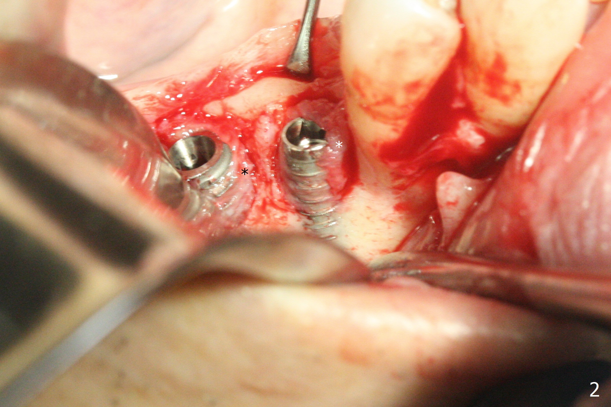
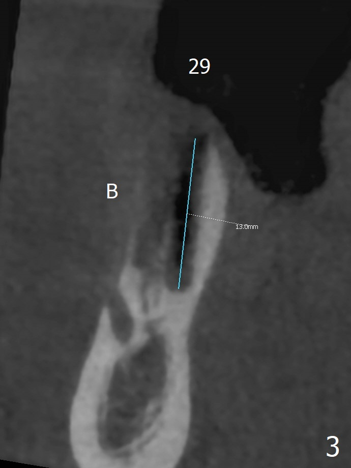
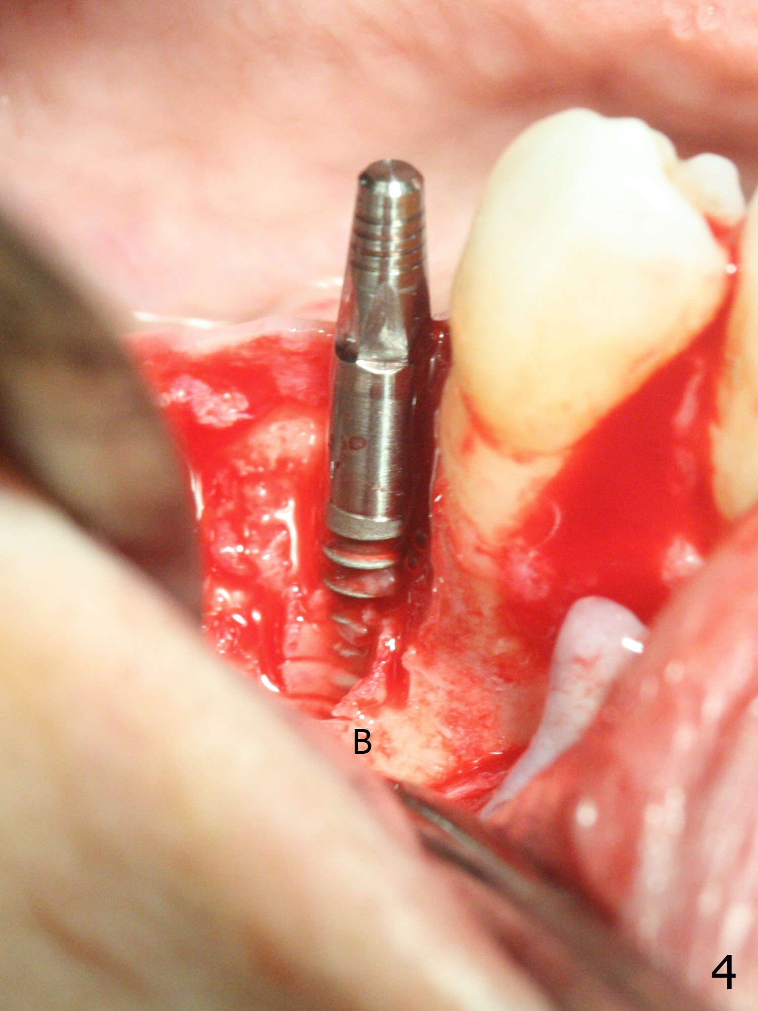
The buccal gingiva is recessive at #29 and 30 (Fig.1). Incision reveals buccal implant thread exposure with circum-ferential granulation tissue (Fig.2 *). Immediate post implant removal at #29, osteotomy is initiated lingual (Fig.3 (intraop CT, coronal section) ). A smaller and shorter implant (3x12 mm vs. 3.5x13 mm) is placed away from buccal (Fig.4 B, 5).
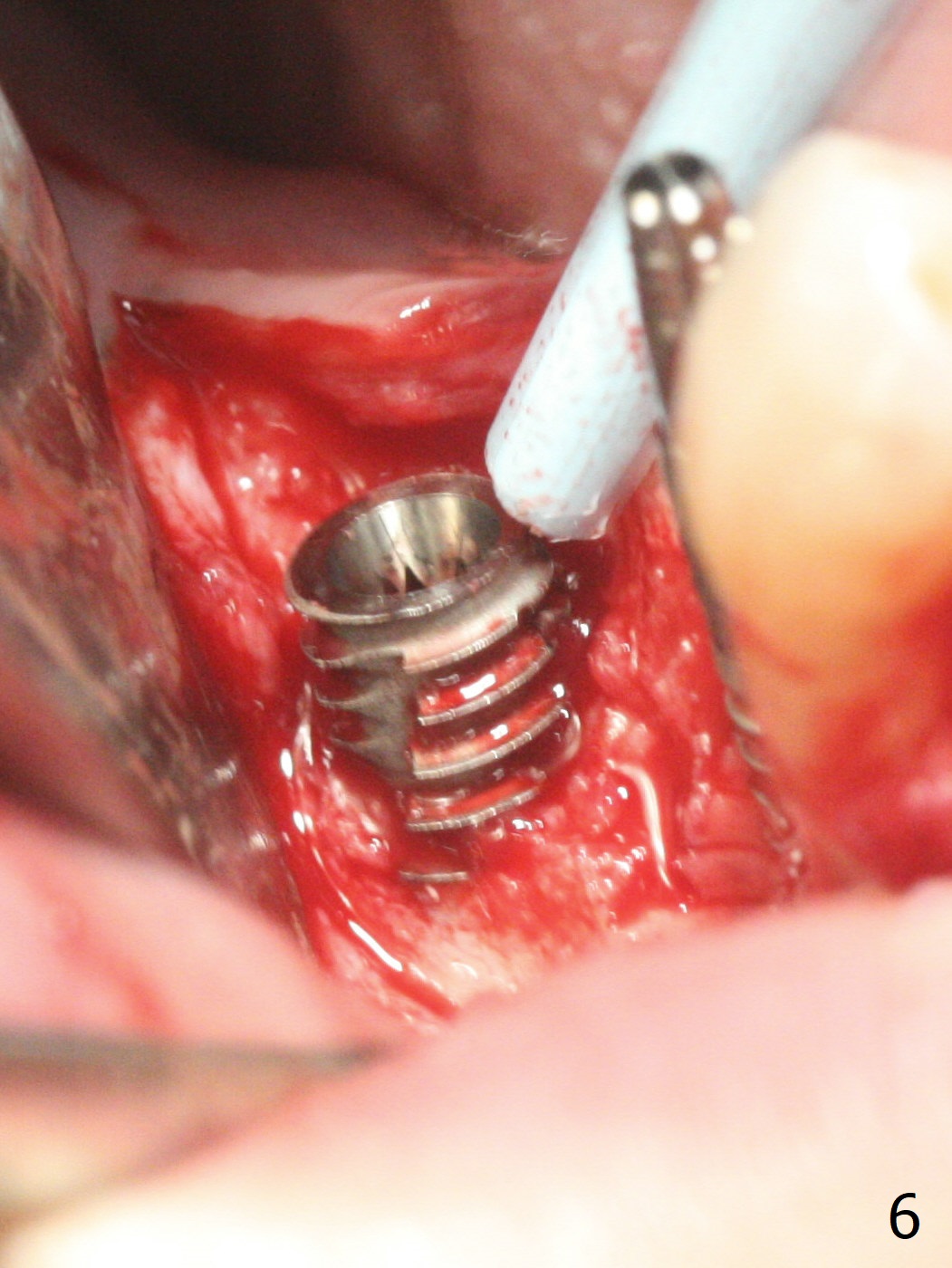
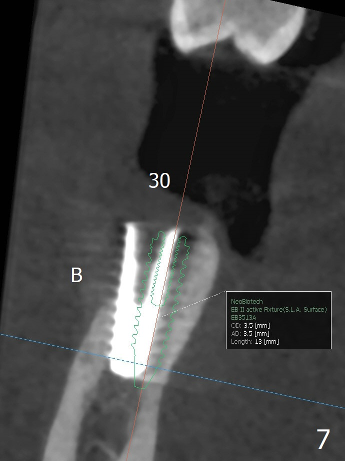
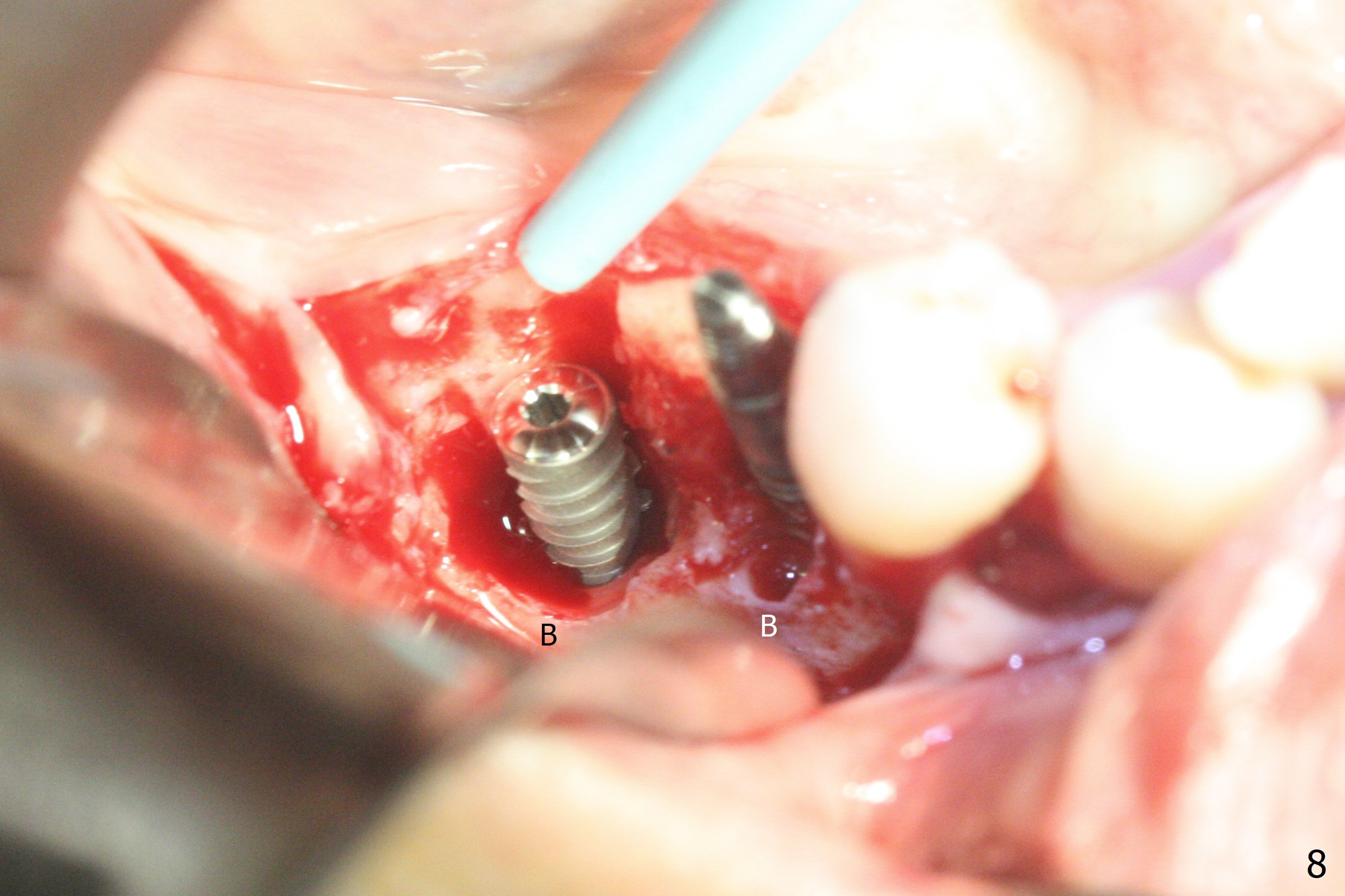
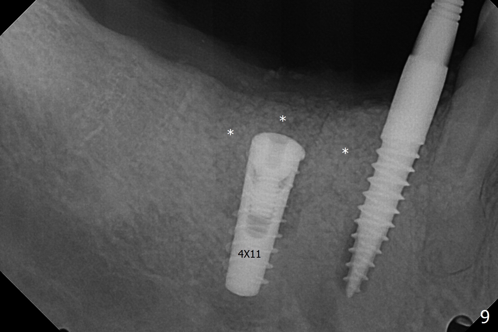
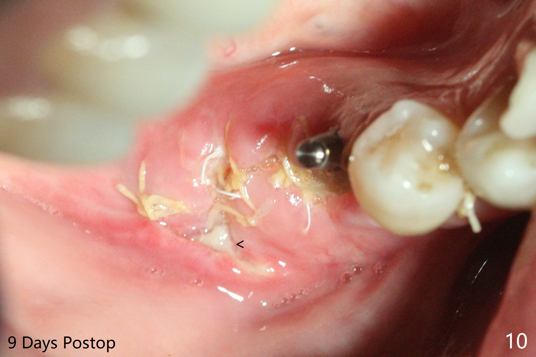
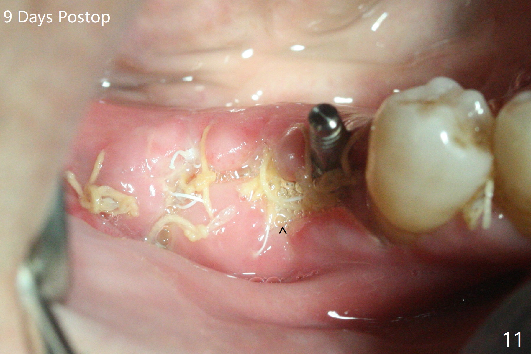
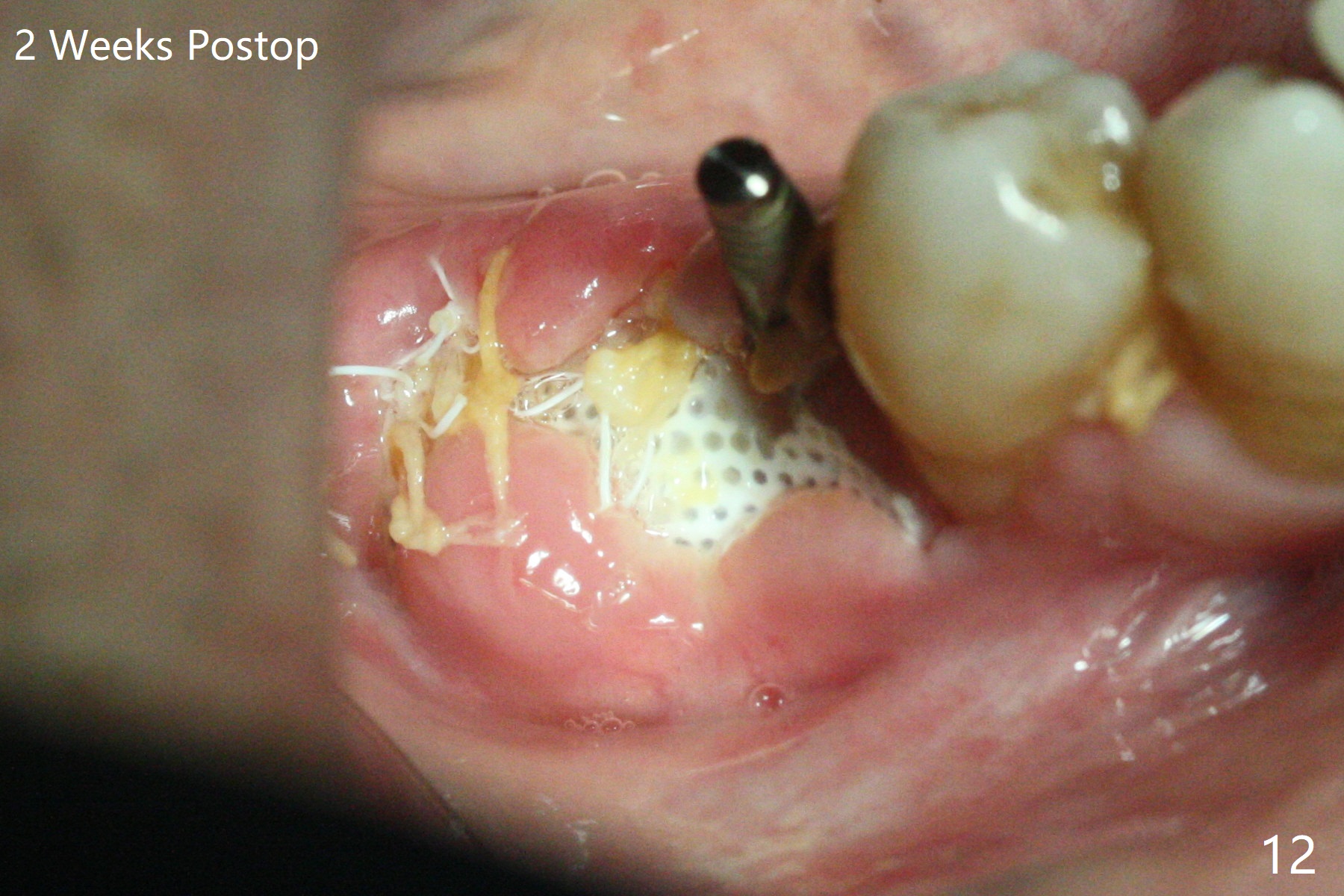
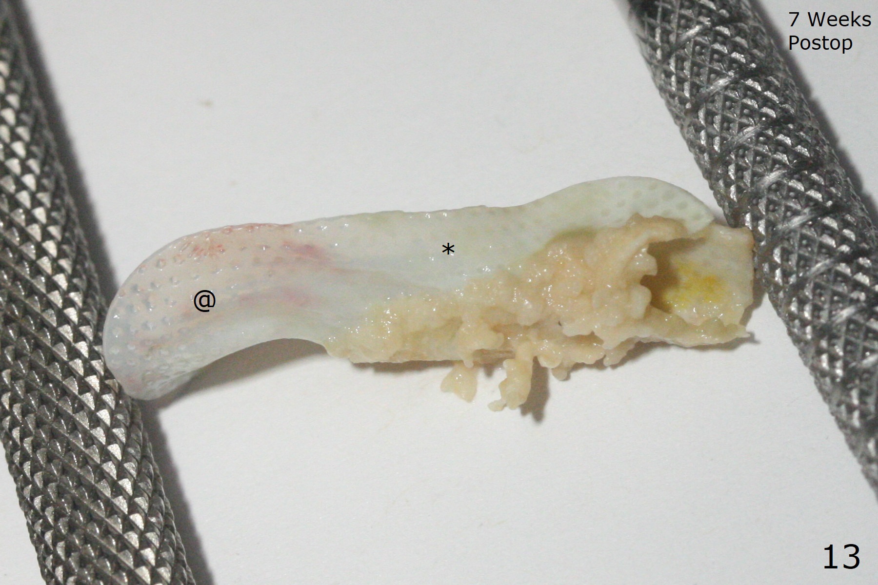
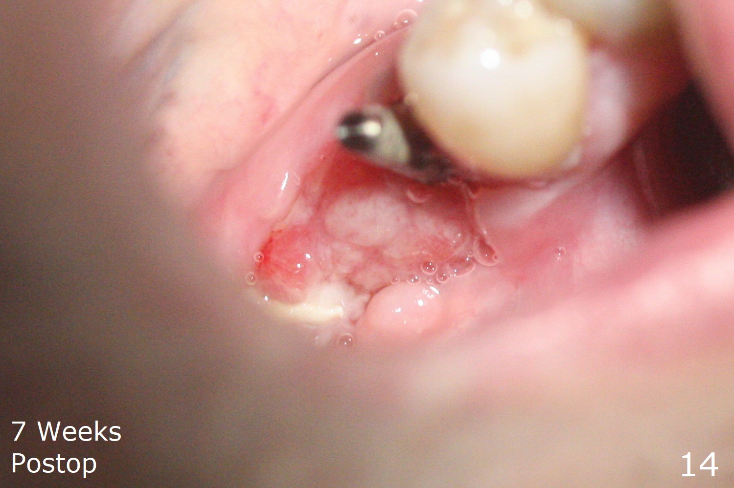
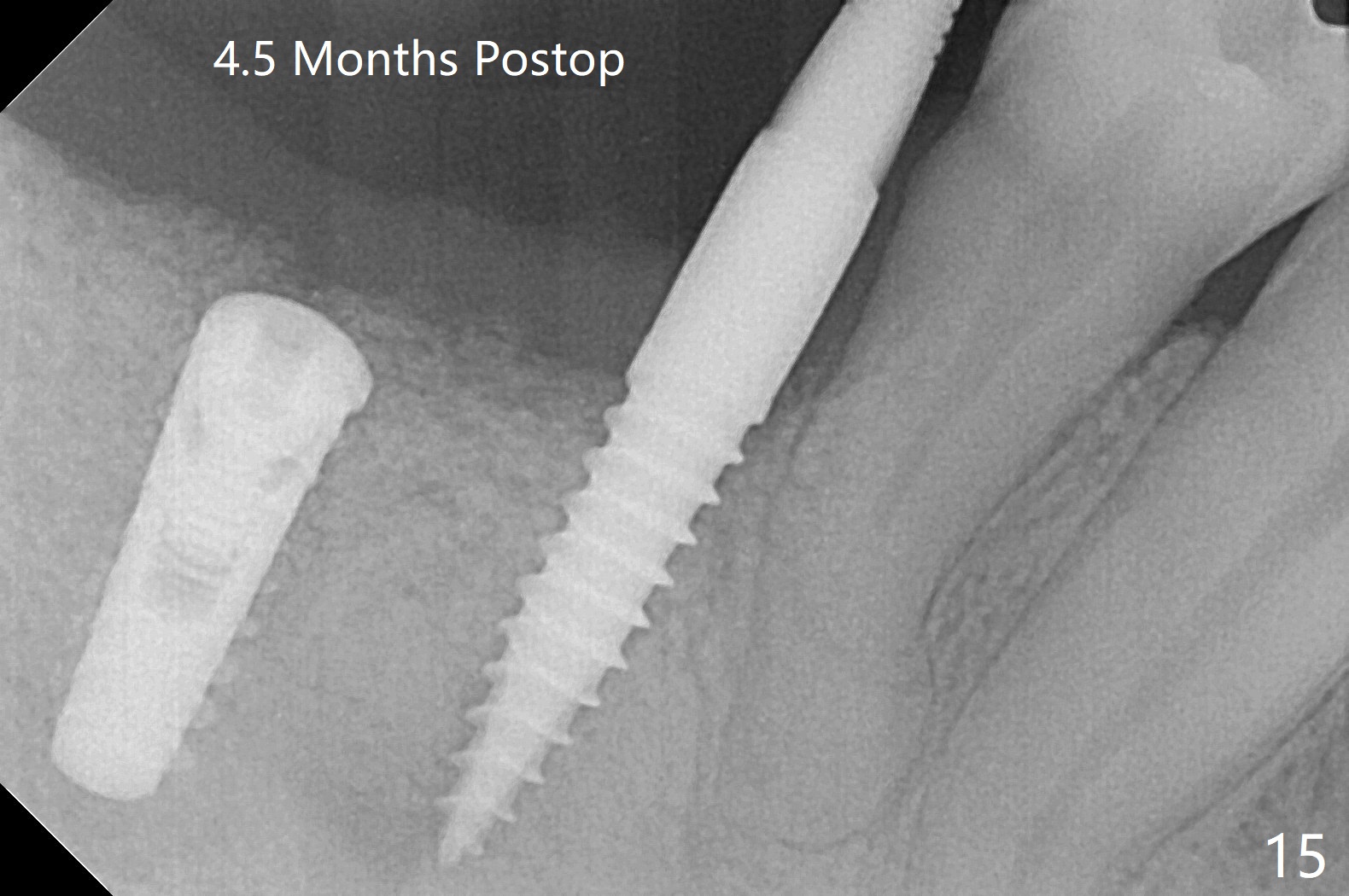
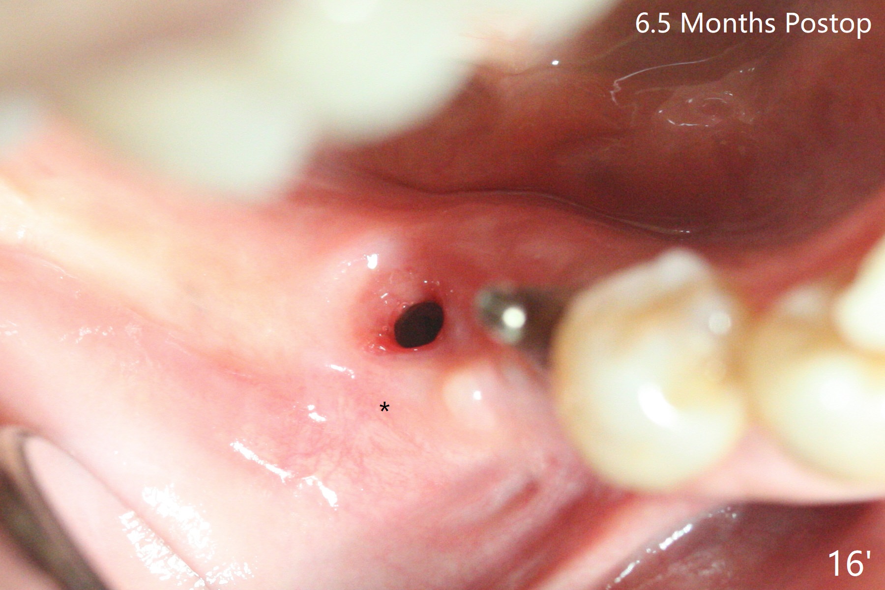
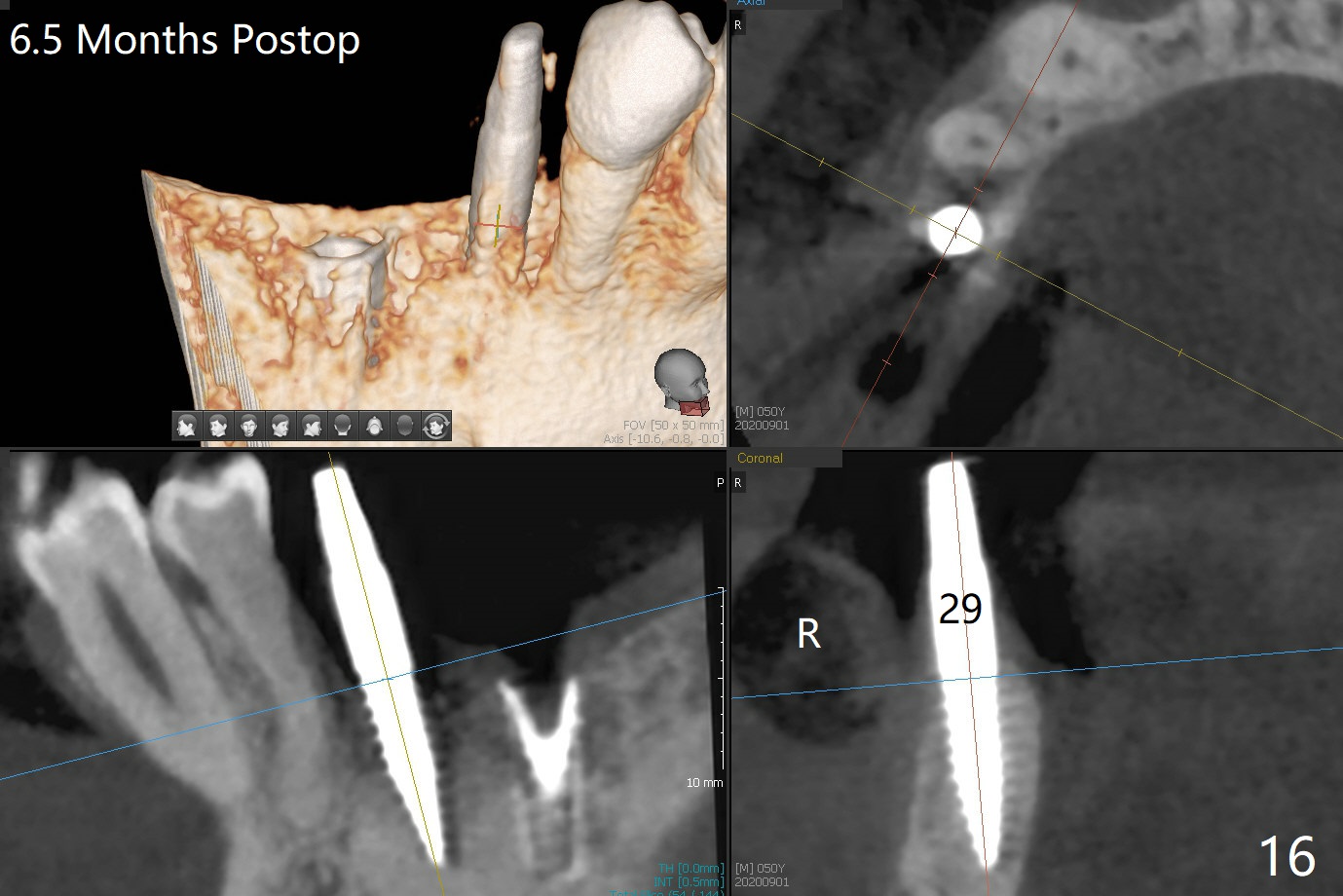
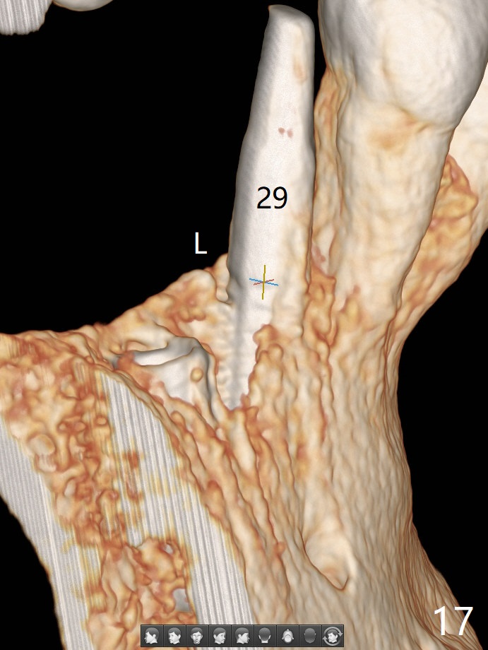
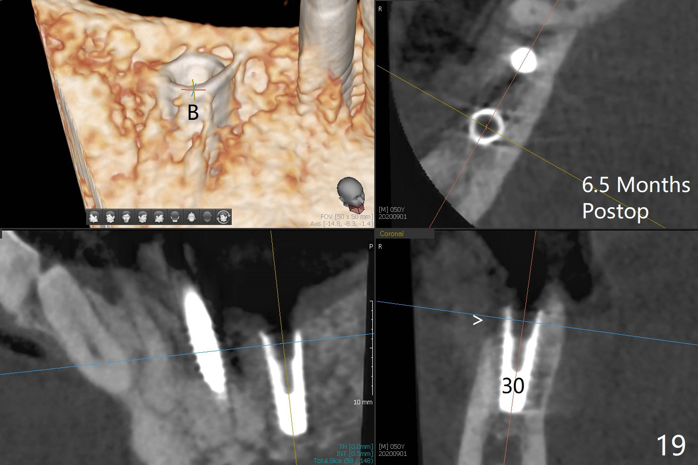
The implant at #30 was also buccally placed (Fig.6,7) and should be corrected in the same manner (Fig.7 green). Due to the bone being harder in the molar region, a smaller and shorter implant (4x11 mm vs. 5x13 mm) shifts slightly buccally while being placed (Fig.8,9). Since primary stability is lower (<20 Ncm vs. 35 Ncm associated with the implant #29), an abutment is not placed, which may be favorable to healing, but it is difficult to achieve primary closure. After bone graft (Fig.9 *) and 2 layers of PRF, Cytoplast is placed. Cytoplast appears to be exposed buccally (Fig.10 <) and occlusally (Fig.11 ^) asymptomatic 9 days postop. Exposure of Cytoplast is more distinct without sign of infection 15 days postop (Fig.12). The patient returns with chief complaint of "foul smell" 7 weeks postop (coronavirus lockdown). Although the Cytoplast exposes more (Fig.13 (* exposed; @ unexposed)), the underlying gingiva remains healthy (Fig.14). While the bone height decreases at #29, the bone density at #30 increases 4.5 months postop (Fig.15). The gingiva heals. The implant at #30 is uncovered 6 months postop. The lingual plate has to be removed for the uncover, while the coronal end of the buccal one is missing. No bone graft is added. When the 4.5x4 mm healing abutment is removed 6.5 months postop, the buccal plate looks concave at #30 (Fig.16' *). The buccal plate looks thin at #29 with a cotton roll placed buccally (Fig.16 R). The lingual plate at #29 is coronal to the buccal one (Fig.17). The buccal gingiva at #29 is quite long (Fig.18). The coronal buccal plate appears to be missing (Fig.19 >), which will be watched. A 4.5x7.5(4) mm cemented abutment is torqued (Fig.20).
Return to
No Deviation Next Case
for
Prevention
Xin Wei, DDS, PhD, MS 1st edition
02/18/2020, last revision
09/01/2020