,%203x10(2)%20d%20after%20E2.jpg)
,%20PRF,%20Vanilla.jpg)
.jpg)
|
|
|
|
|
|
|
|
|
|
|
|
|
|
Different Type, Size and Length of Implants
Depending On Bone Variation
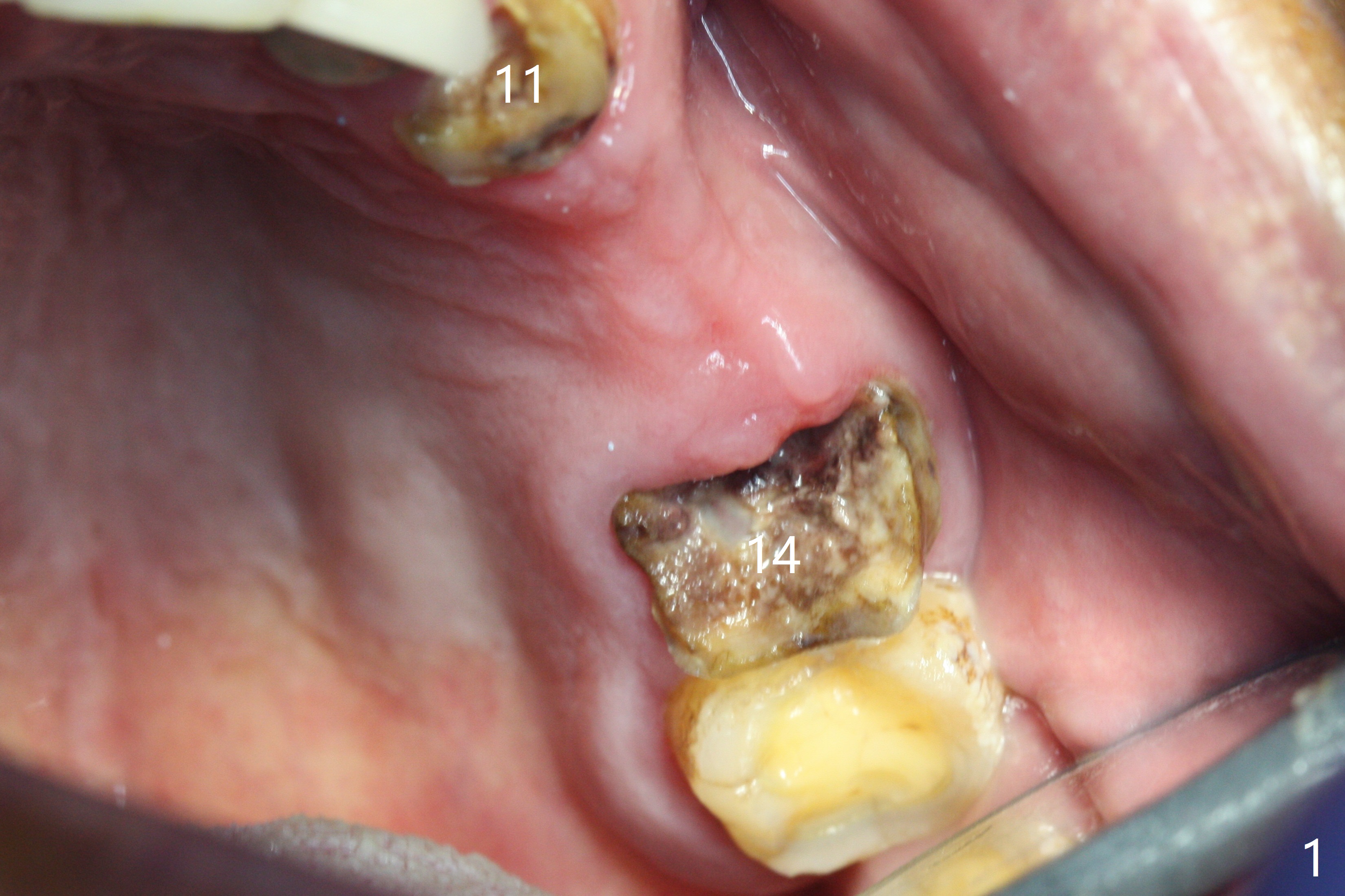
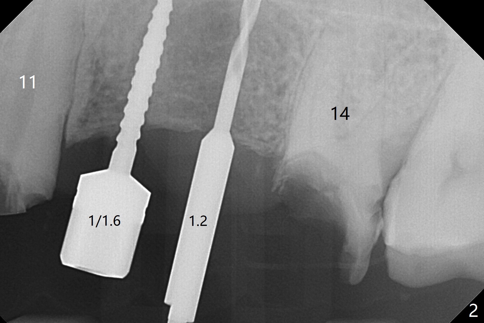
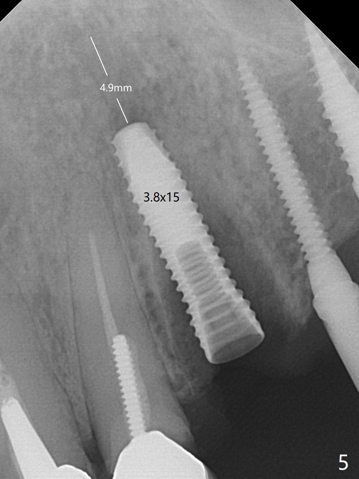
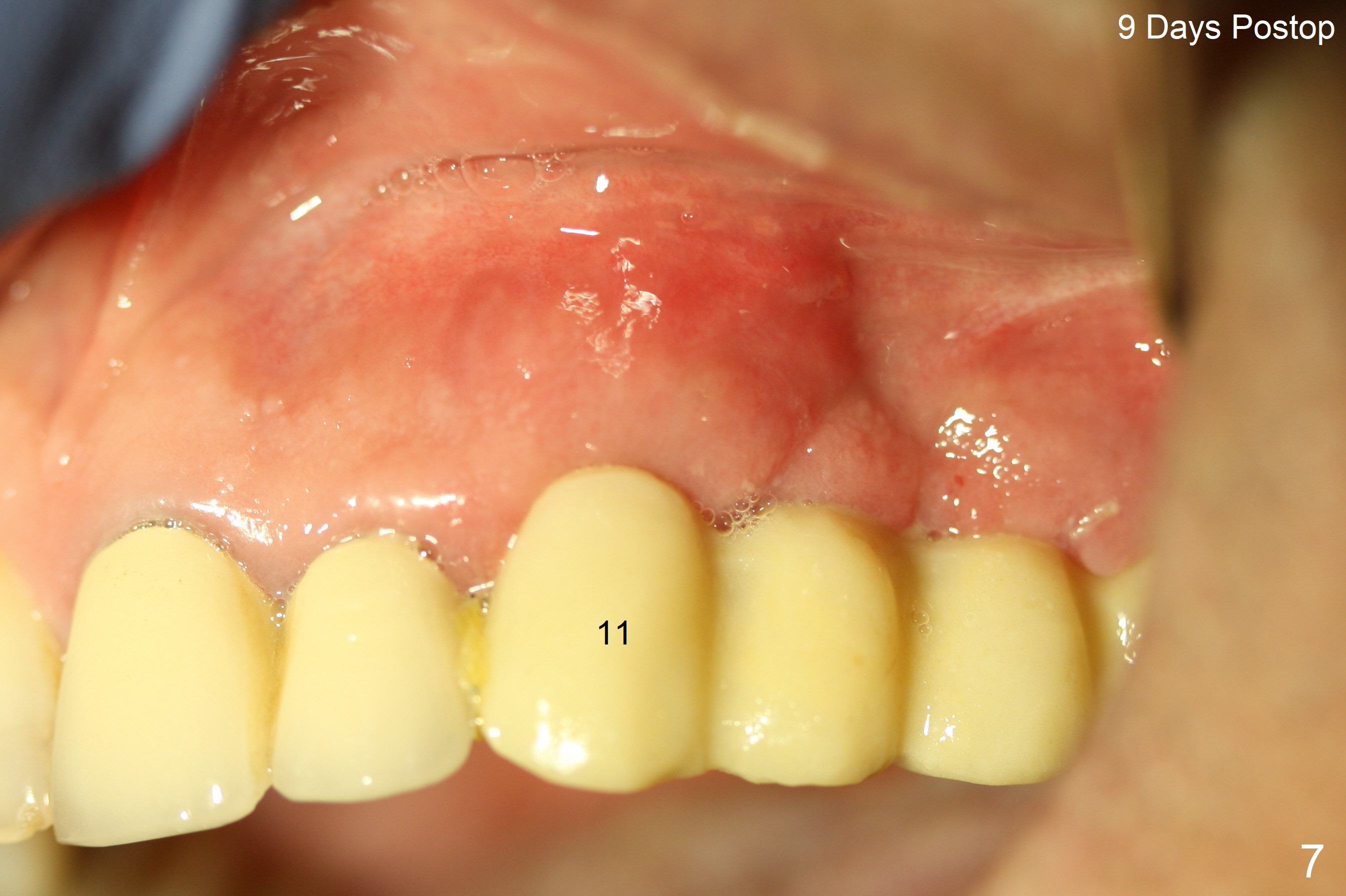
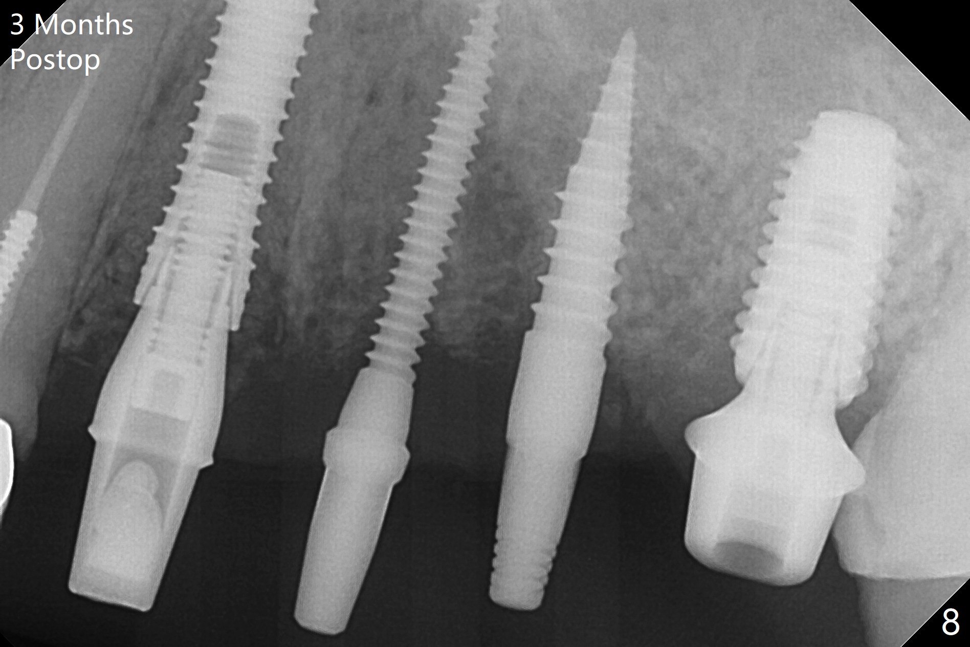
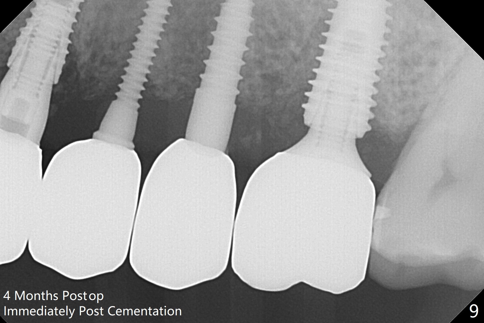
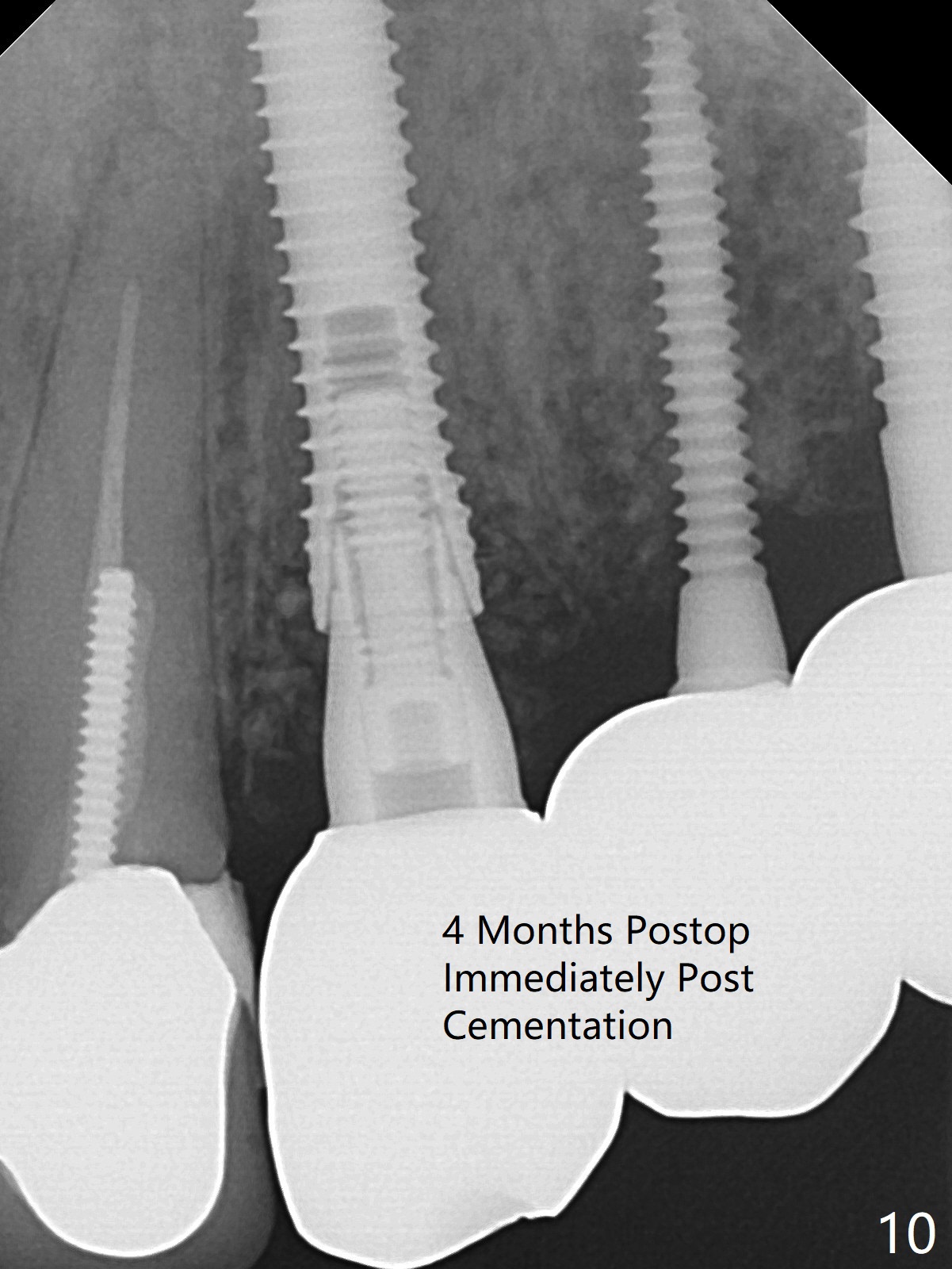
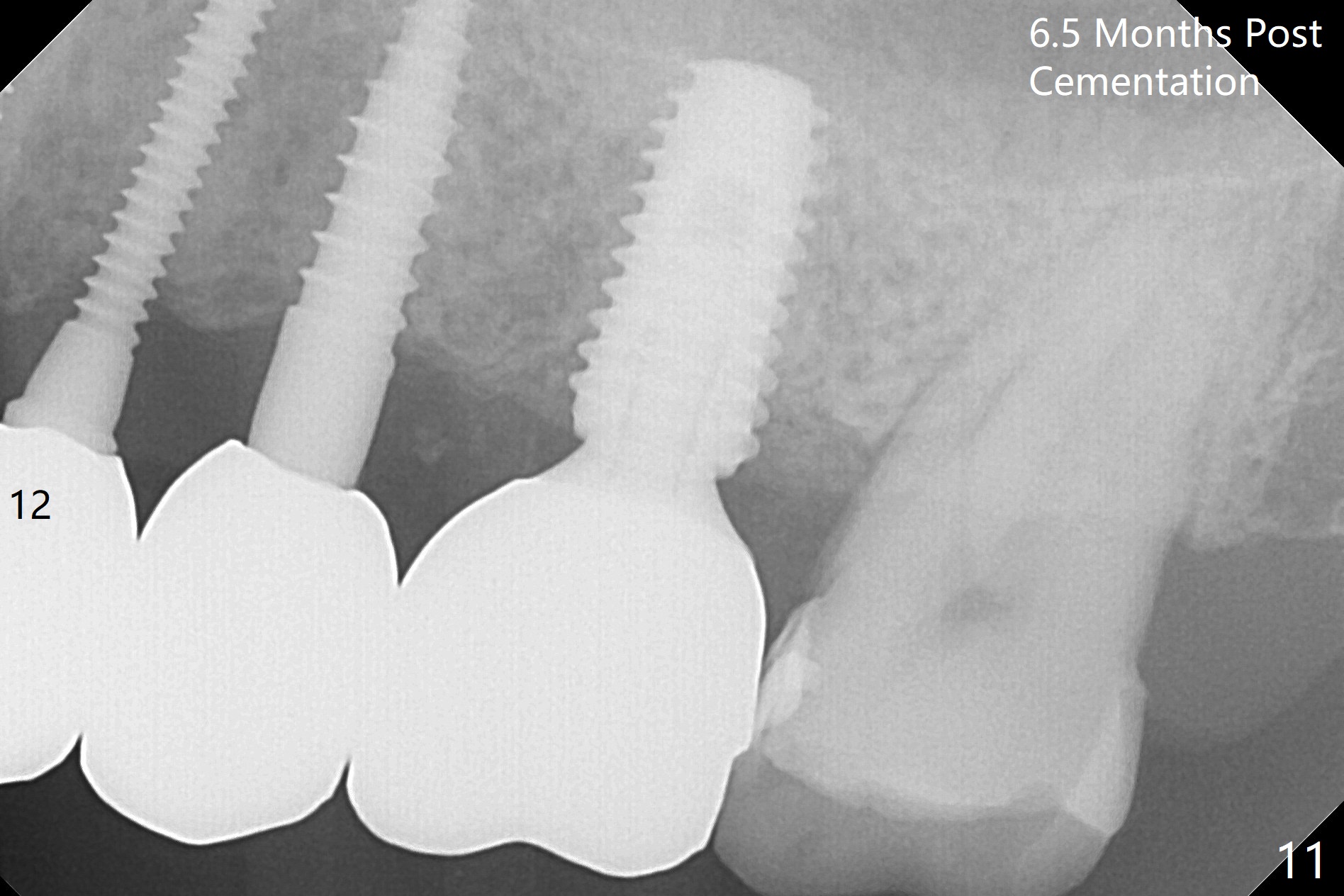
The edentulous ridge is narrow between the residual roots of #11 and 14 (Fig.1). Since the bone density is low in the edentulous area, DIO bone expanders are used (e.g., #1 (1/1.6 mm) Fig.2) after 1.2 mm initial drill. A 2x14(2) mm 1-piece implant is placed at #12 with 4 mm ridge width, while a 3x10 (2) mm dummy implant is partially placed after use of Bone Expander #2 (1.3/2.3 mm). The latter is replaced by a definitive one (3x12(2) mm), while the one at #12 is placed deeper (Fig.4). After extraction and placement of PRF and Vanilla Graft for sinus lift (Fig.4 black *), a 5x10 mm 2-piece implant is placed at #14, while a dummy implant is placed at #11 (Fig.4,5). As the osteotomy at #11 is 4.9 mm longer than the dummy (Fig.5), a definitive one is 3 mm longer (Fig.6). Osteogen plug is placed in the apical portion of each socket at #11 and 14, while Vanilla and Osteogen are packed in the coronal portion of the socket (Fig.4,6 *). Although primary stability of each implant is not high (30/40 Ncm), splinted provisional with occlusal clearance seems to be sufficient for implant osteointegration. There is no discomfort 9 days postop (Fig.7). Impression is taken 3 months postop, since he plans to return to home country for business (Fig.8). Single unit crowns are cemented 4 months postop (Fig.9,10). The abutment screw at #14 needs retightening 6 months post cementation. The access hole slightly buccal. Chewing pain at #12 is reduced after occlusal adjustment 6.5 months post cementation (Fig.11).
Return to Upper Canine, Molar Immediate Implant, Prevent Molar Periimplantitis (Protocols, Table), Armaments Xin Wei, DDS, PhD, MS 1st edition 05/08/2018, last revision 03/21/2019