|
|
|
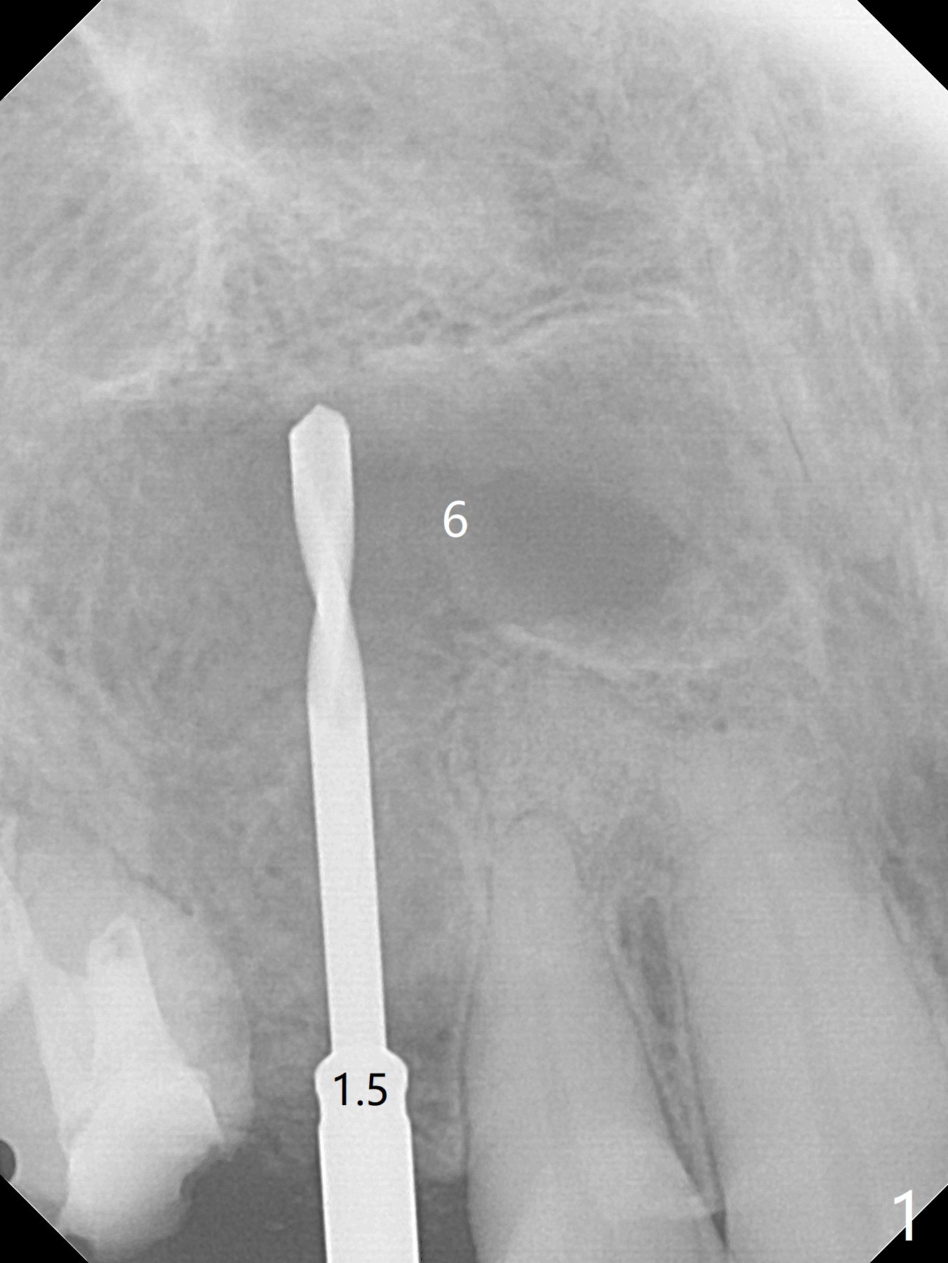
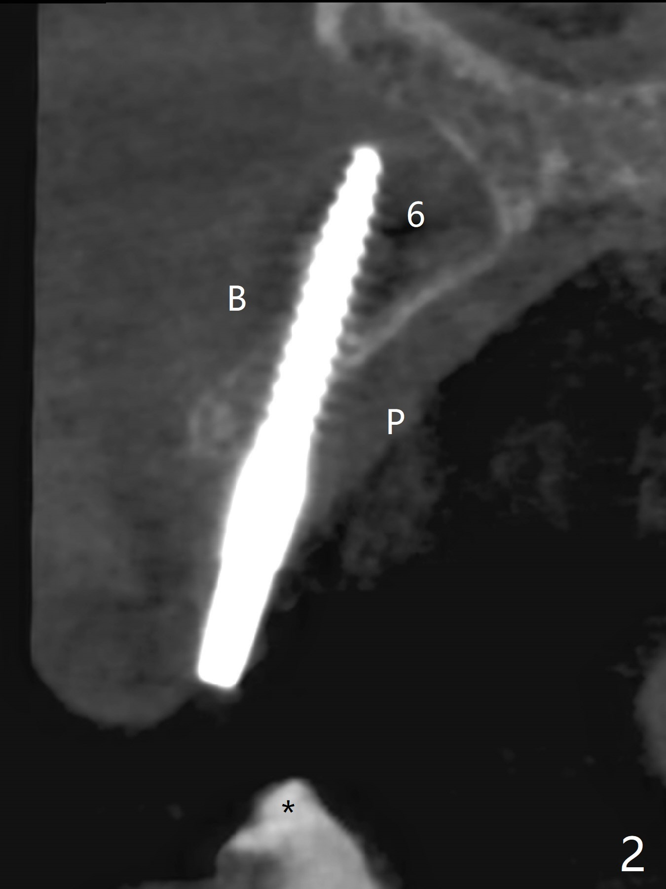
Osteotomy starts with guide and 2.2 mm drill for initial 3.5 mm palatal. A semilunar incision is made buccal to remove the impacted canine (#6 by sectioning). With direct vision buccal and palatal, the final osteotomy is finished free hand with 1.2 and 1.5 mm drills. Initially buccal perforation occurs, the osteotomy route is corrected later (Fig.1). When a 2.5x15 mm 1-piece implant is being placed, it perforates into the buccal concavity again. After redirection, the trajectory seems to be acceptable (Fig.2: CT coronal section).
Xin Wei, DDS, PhD, MS 1st edition 01/14/2019, last revision 01/29/2019