|
|
|
|
|
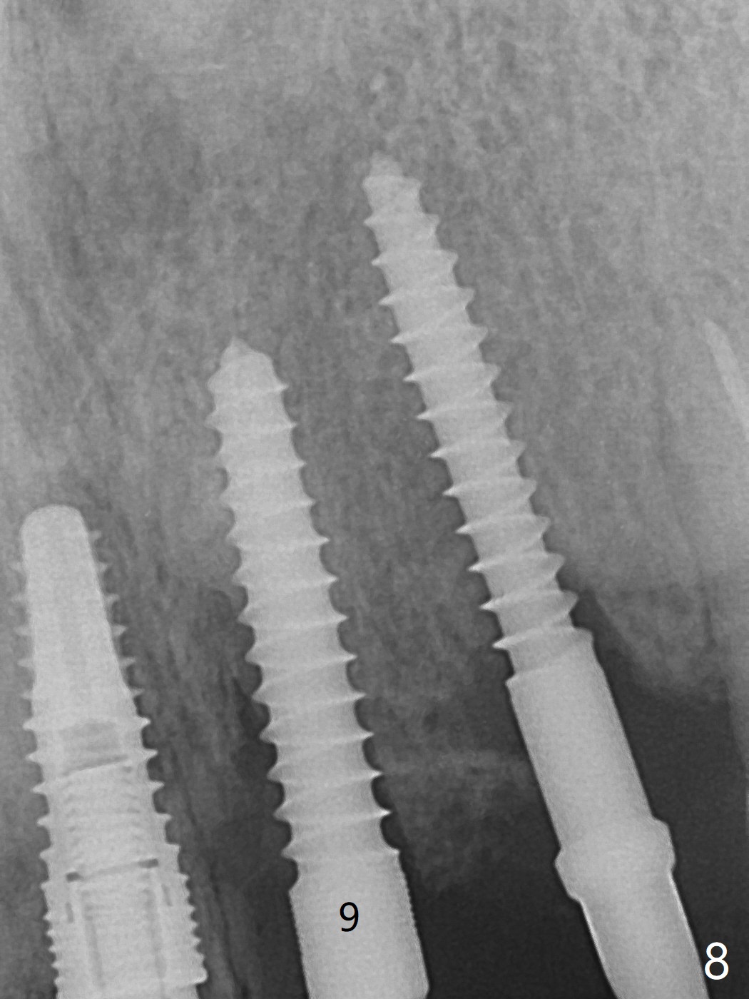
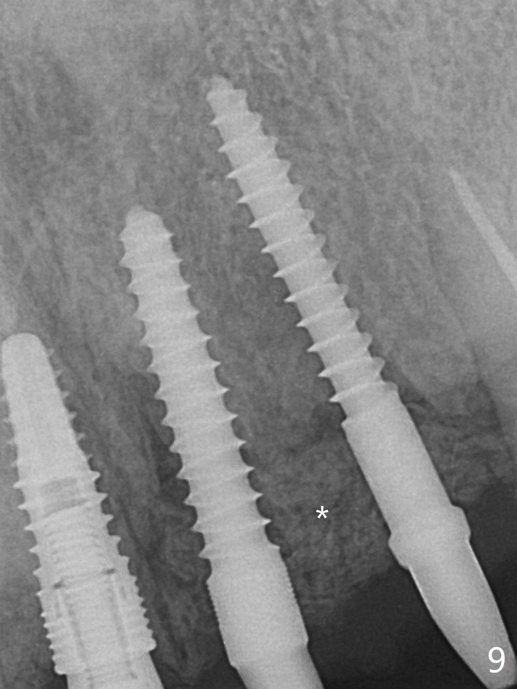
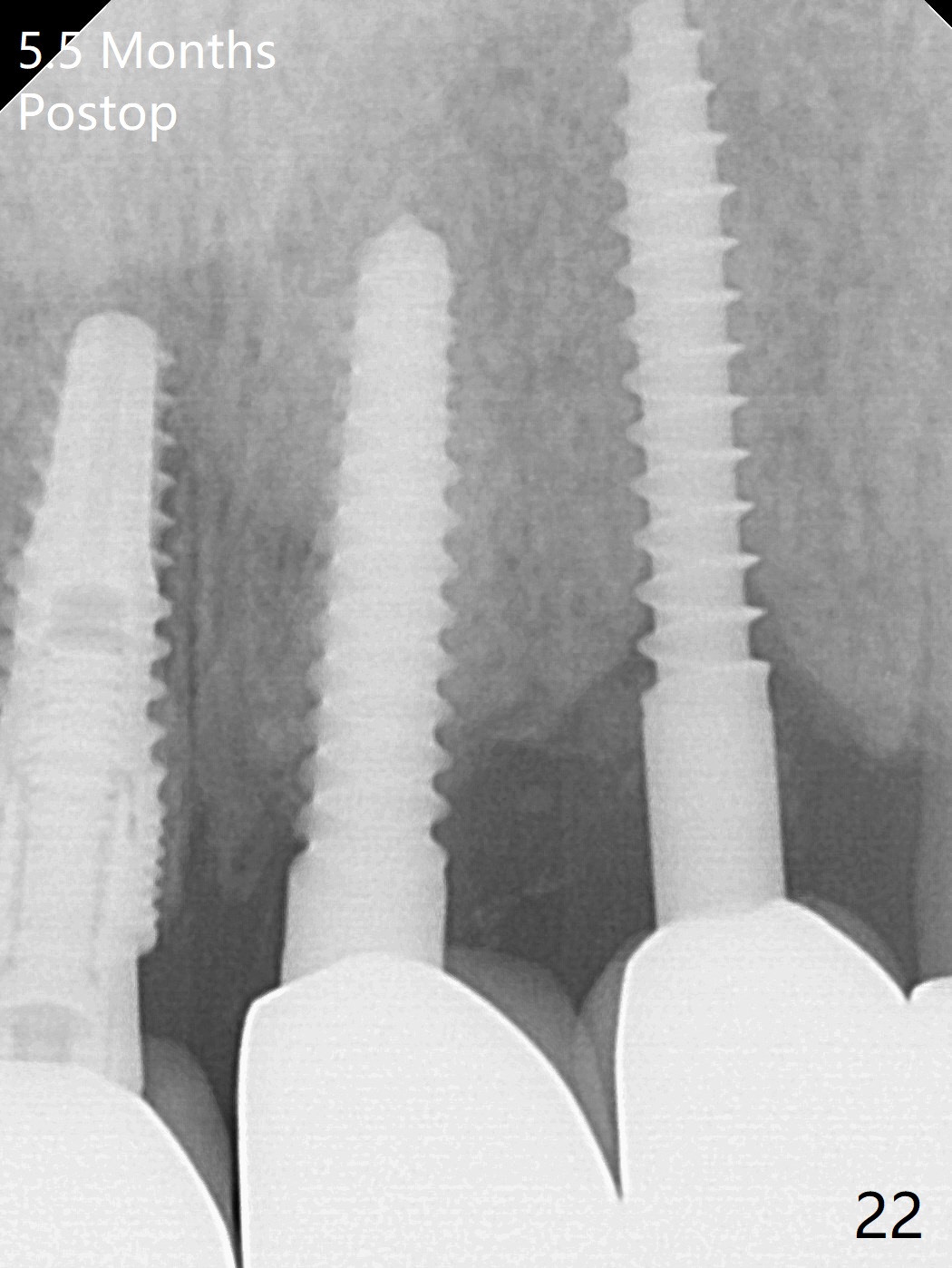
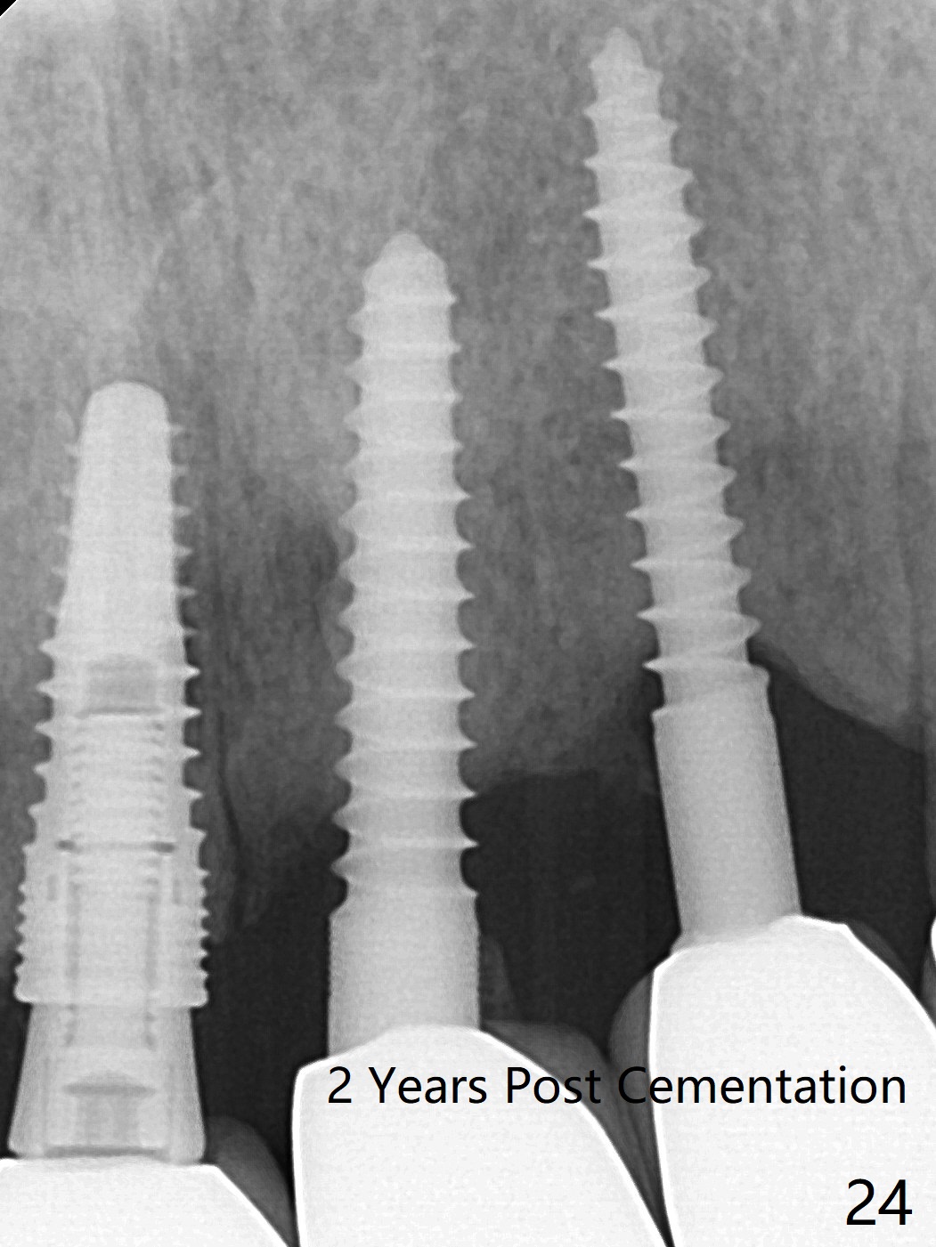
After re-adjustment (Fig.7), the implant is placed at the right orientation (Fig.8). Allograft is placed mainly buccally (Fig.9,10 *), followed by a piece of collagen membrane (Fig.11). After tension release, flaps are approximated (Fig.12). There appears to be new bone formation around the coronal implant threads 5.5 months postop (immediately post cementation, Fig.22). The microthreads at #9 may be not bone covered, the reason for the gingival erythema.粘固后拍摄根尖片检查残余粘固剂。粘固后两年牙槽嵴骨质并没有再生(图二十四),说明第一术中植体必须植入骨下(基台部分要长,否则难于修复),第二牙槽嵴处不应该有压力,植入2.5毫米植体,最后钻头应该是2.5毫米,骨下1-3毫米(尝试项目)。
Palatal Placement Last Next 位点保存
Xin Wei, DDS, PhD, MS 1st edition 07/02/2021, last revision 07/04/2021