|
|
|
|
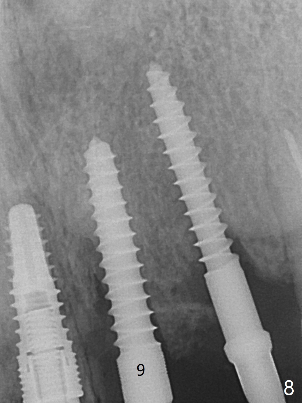
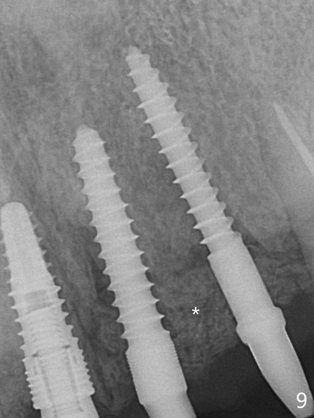
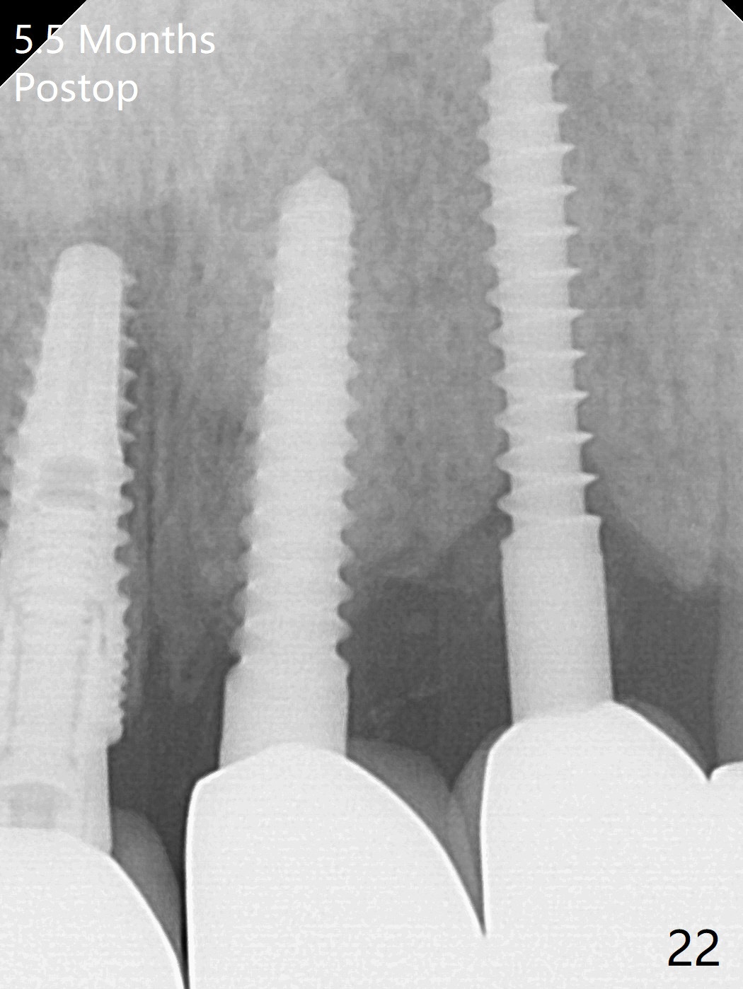
After re-adjustment (Fig.7), the implant is placed at the right orientation (Fig.8). Allograft is placed mainly buccally (Fig.9,10 *), followed by a piece of collagen membrane (Fig.11). After tension release, flaps are approximated (Fig.12). There appears to be new bone formation around the coronal implant threads 5.5 months postop (immediately post cementation, Fig.22). The microthreads at #9 may be not bone covered, the reason for the gingival erythema.粘固后拍摄根尖片检查残余粘固剂。
Palatal Placement Last Next Xin Wei, DDS, PhD, MS 1st edition 07/05/2019, last revision 07/04/2021