 |
 |
 |
 |
 |
Thin Bone M
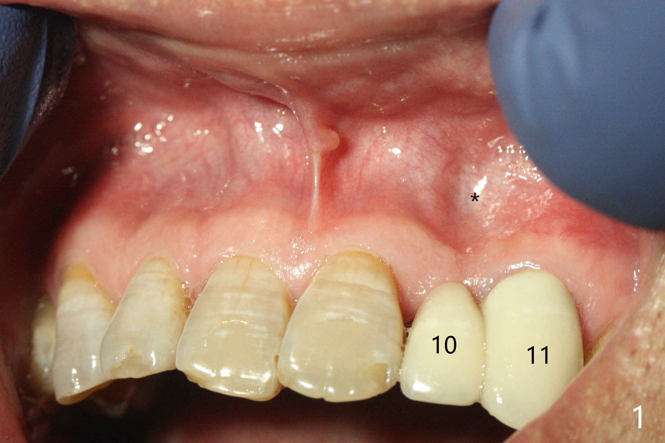
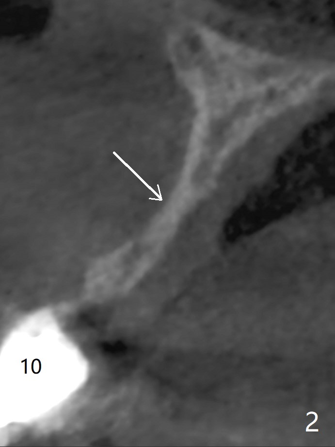
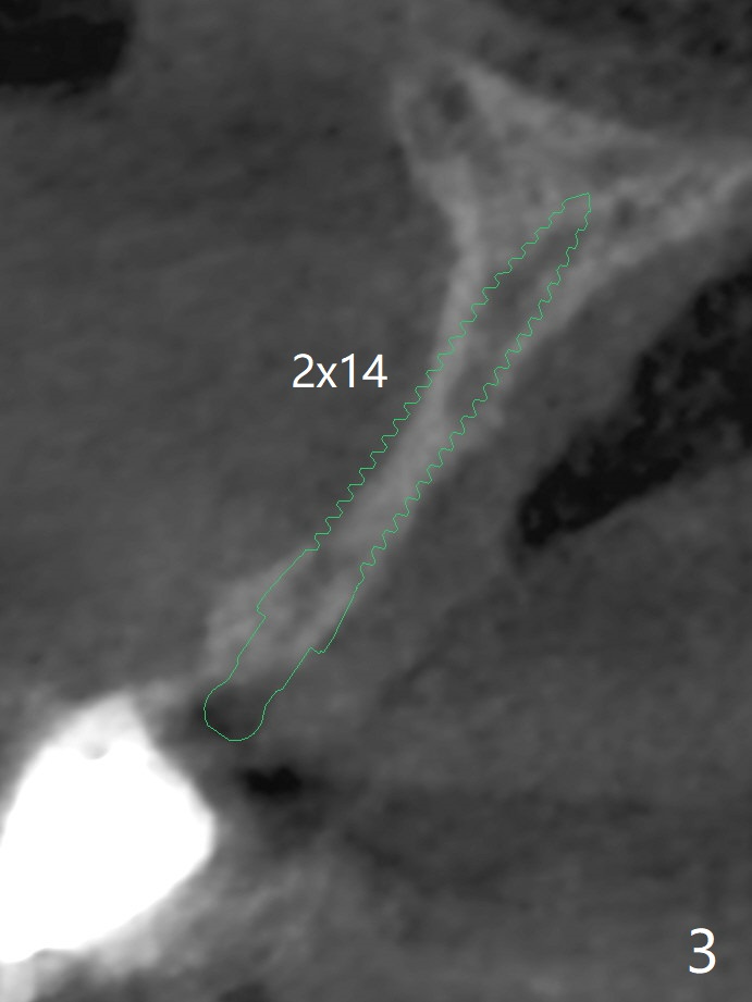
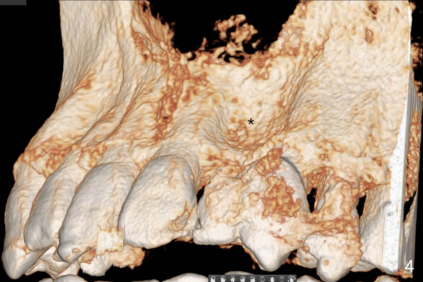
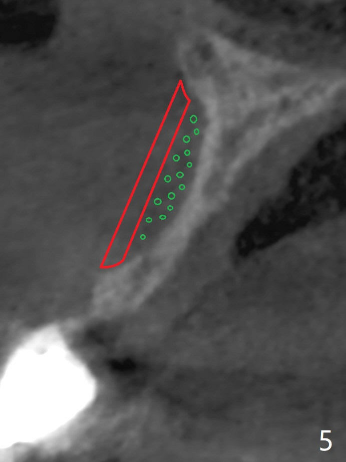
A 46-year-old man requests implant at #10 because of inability to incise on the left (Fig.1). The tooth was extracted 20-30 years since it was malpositioned. There is a severe indentation (Fig.1,4 *). The buccal and palatal plates are fused in the deepest area of the concavity (Fig.2 arrow). The thinnest implant (2 mm in diameter, Fig.3 green) is wider than the ridge. It appears that onlay (Fig.5 red) and particulate (green circles) grafts are needed.
Hello Dr. Wei,
This case can defiantly be done by grafting with Bond Apatite
However, since there is almost no spongy bone we need to revive the area
Therefore we need to inform the patient that it might take 2-3 times to reach the desired volume for implant placement. The advantage is that the procedures are minimally invasive, so its healing experience not supposed to be traumatic.
First, we need to revive the bone by reflecting the flap minimally as we indicate with the protocols, then preform deep decertification, place 0.5cc of 3d bond and close the flap by stretching the flap. Leave it for 4-7 weeks, no more.
Open the flap again and now you will see something similar to granulation tissue which is enriched with blood vessels and cells, don't clean it is gold for the success
Just place on it the Bond Apatite and slightly overfill.
Press and compact properly according to the protocols and immediately stretch and suture the flap firs mesially, then distal, then in the middle and in between.
Make sure not to use fast resorbable sutures and not to use removable provisional.
After 3-4 month you can open for implant placement or additional layer if needed.
Please see the protocols for flap reflection and cement application in the link below
Should you have any additional questions please don't hesitate to contact me directly,
Or by phone or WhatsApp
+972523813316
Best
Amos
Return to
Upper Incisor
Immediate Implant
Trajectory II
No Deviation
Xin Wei, DDS, PhD, MS 1st edition
10/21/2019, last revision
03/03/2020