
 |
 |
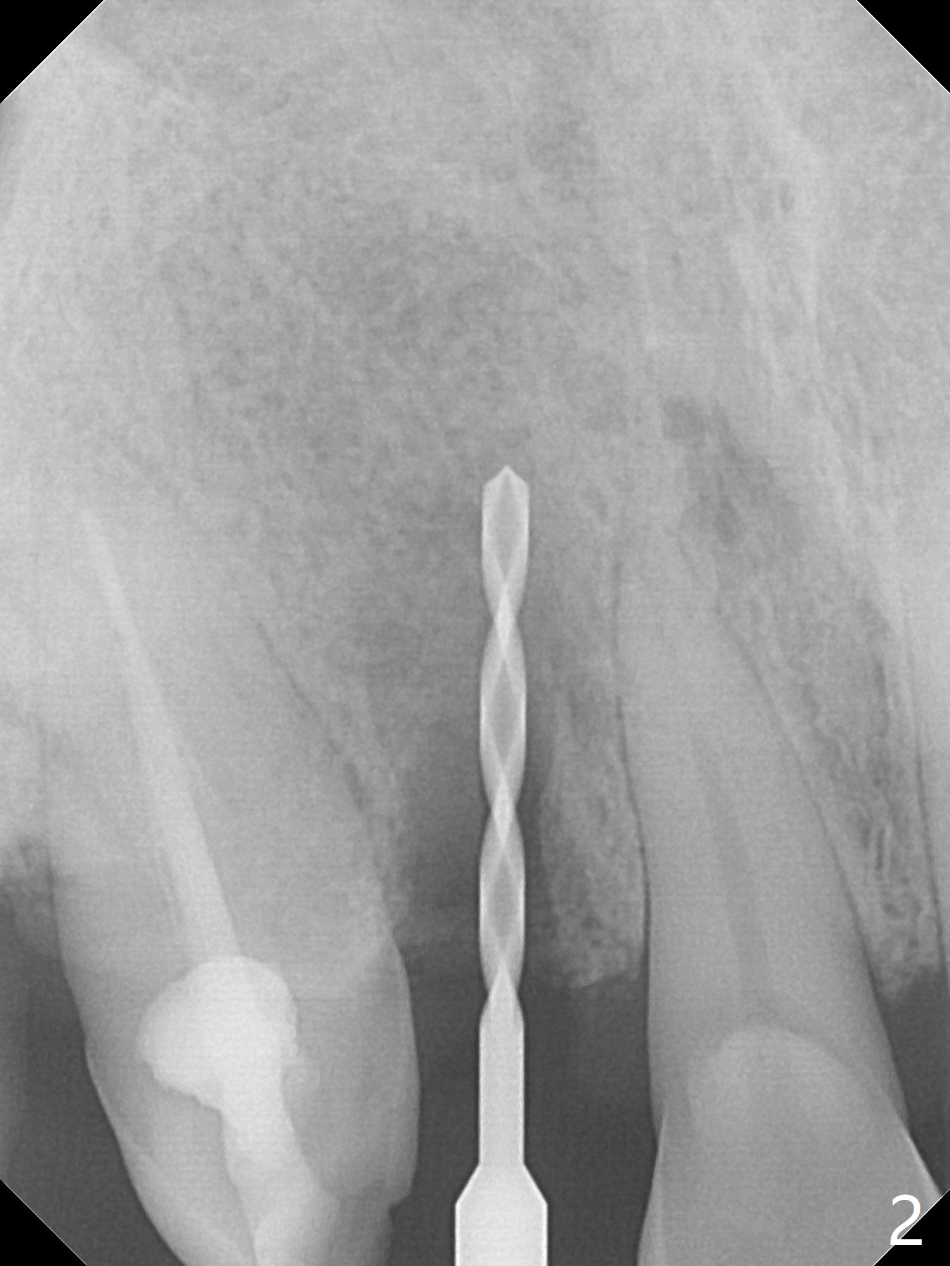
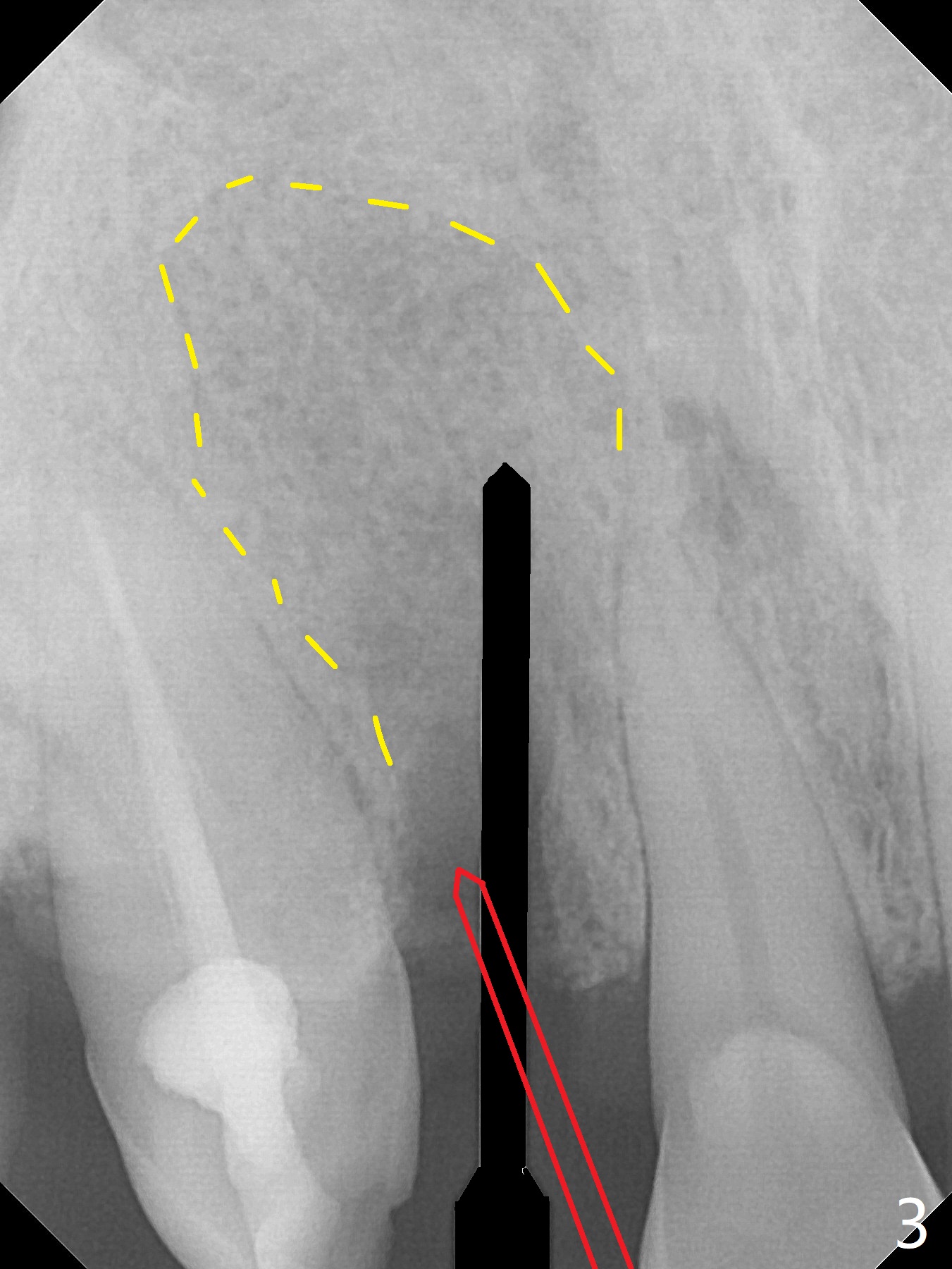
After smooth extraction, the apical buccal plate is found to be perforated. Following debridement, a piece of gauze is placed in the apical defect for hemostasis, while osteotomy is initiated palatal (Fig.2). The apical defect seems to be extensive (Fig.3 yellow dashed line). A new trajectory is intended (red arrow) without much success.
Graft Before Implant Last Next
Xin Wei, DDS, PhD, MS 1st edition 05/07/2019, last revision 03/19/2020