|
|
|
|
|
|
|
|
|
|
|
|
|
|
|
|
|
|
Sinus Lift
with Bone Expanders
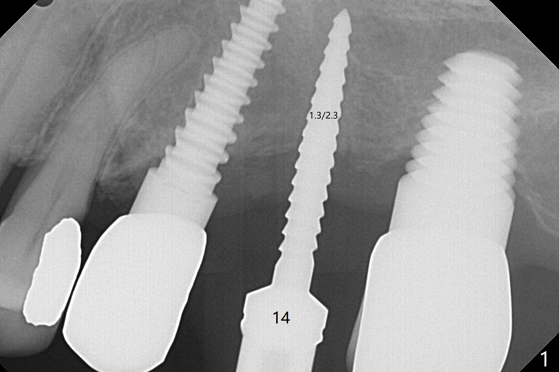
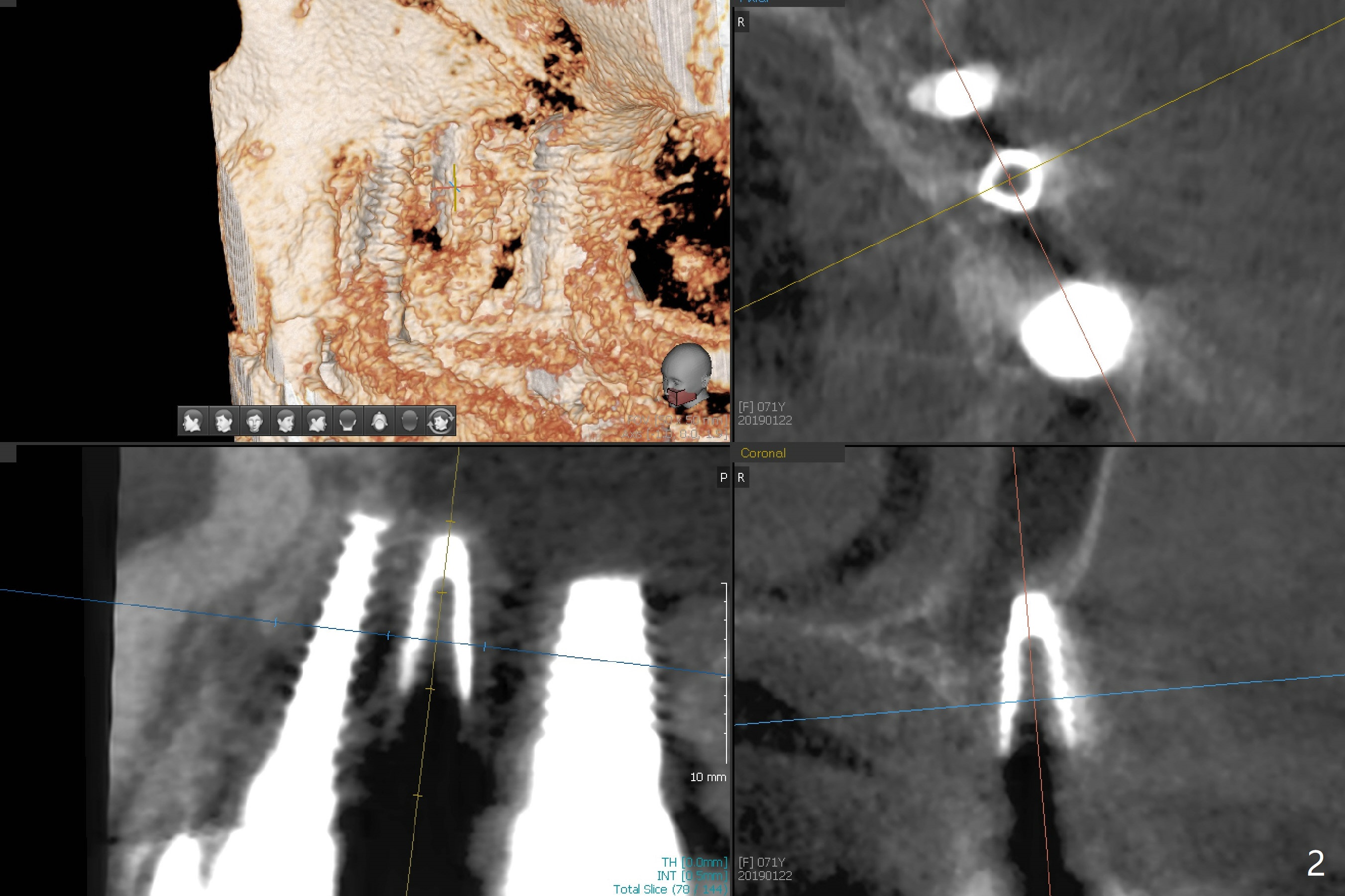
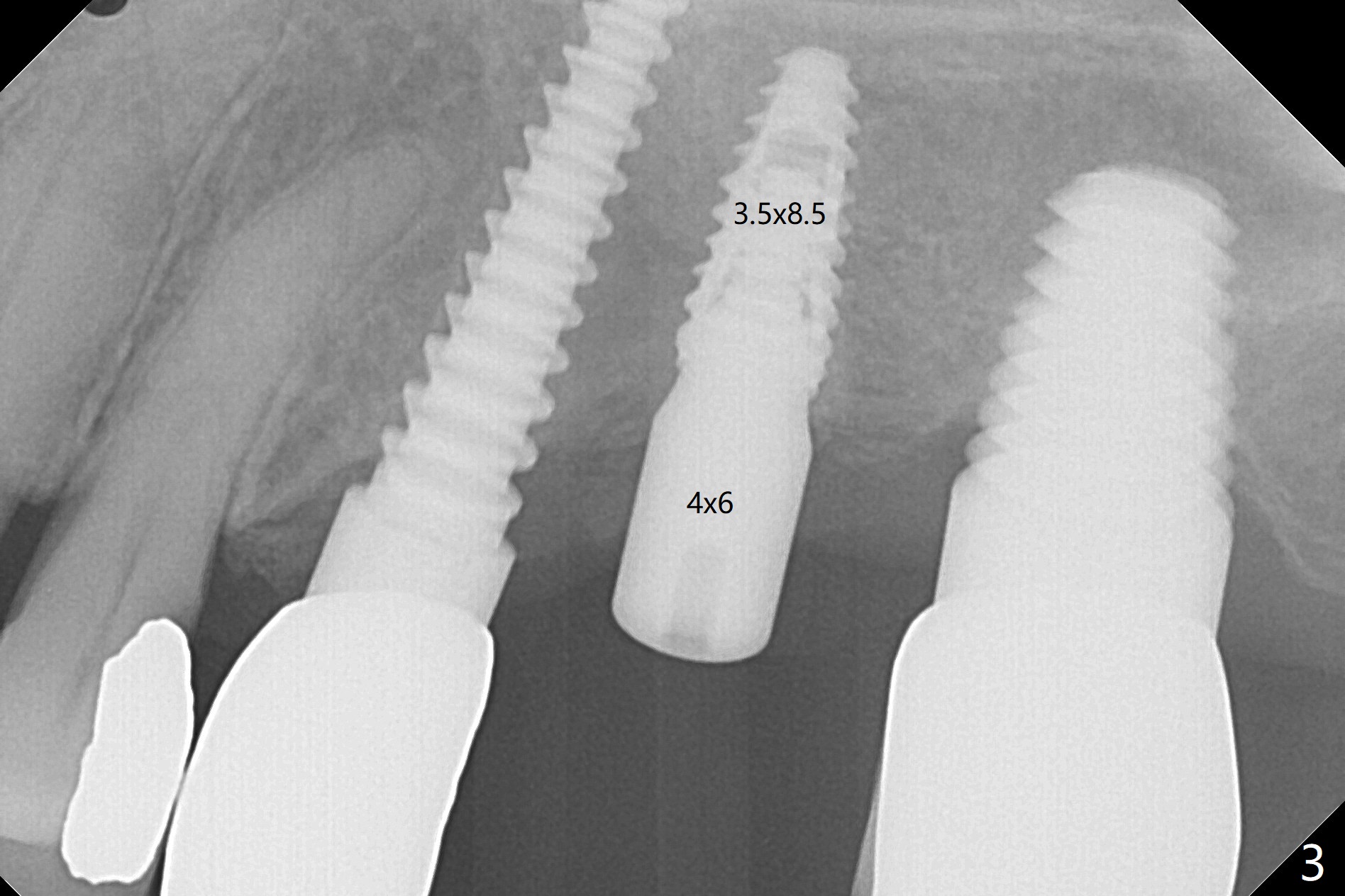
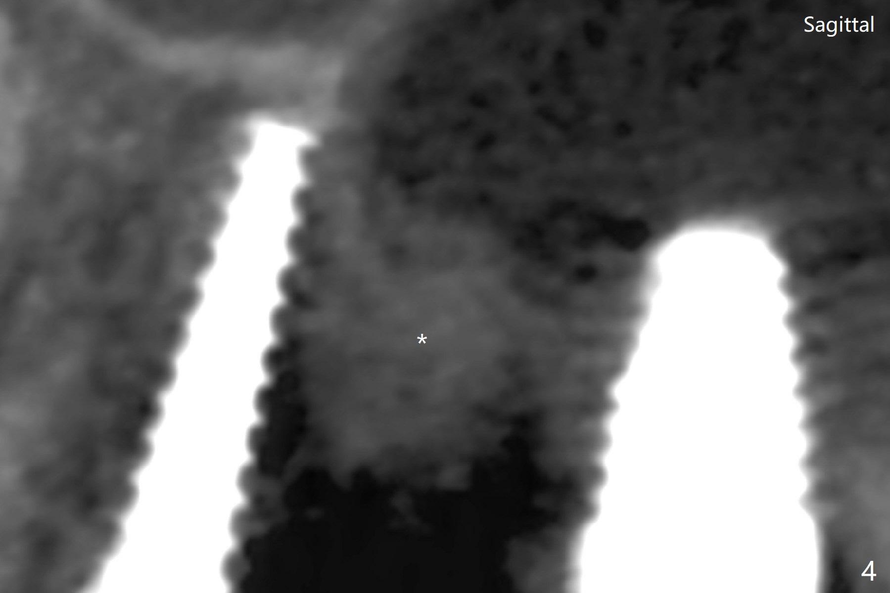
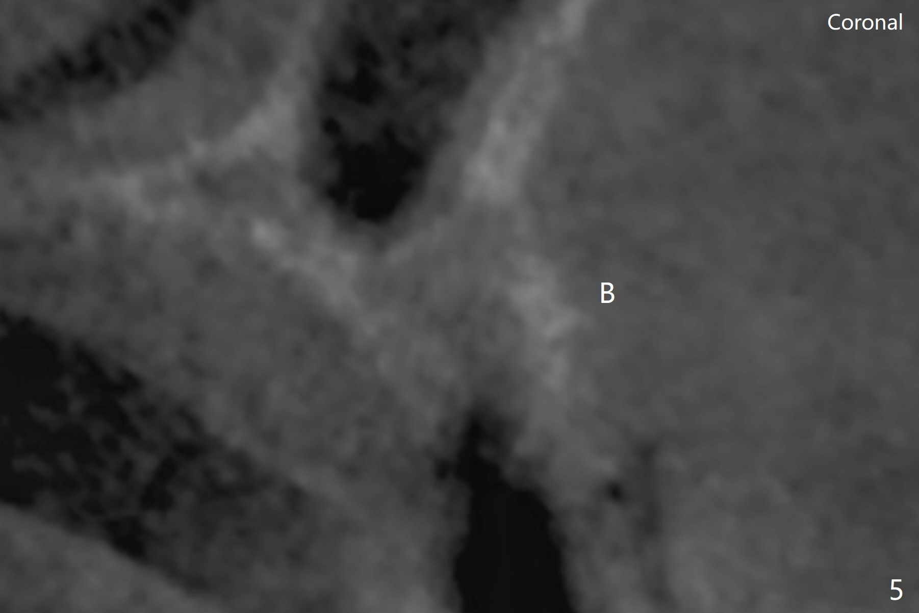
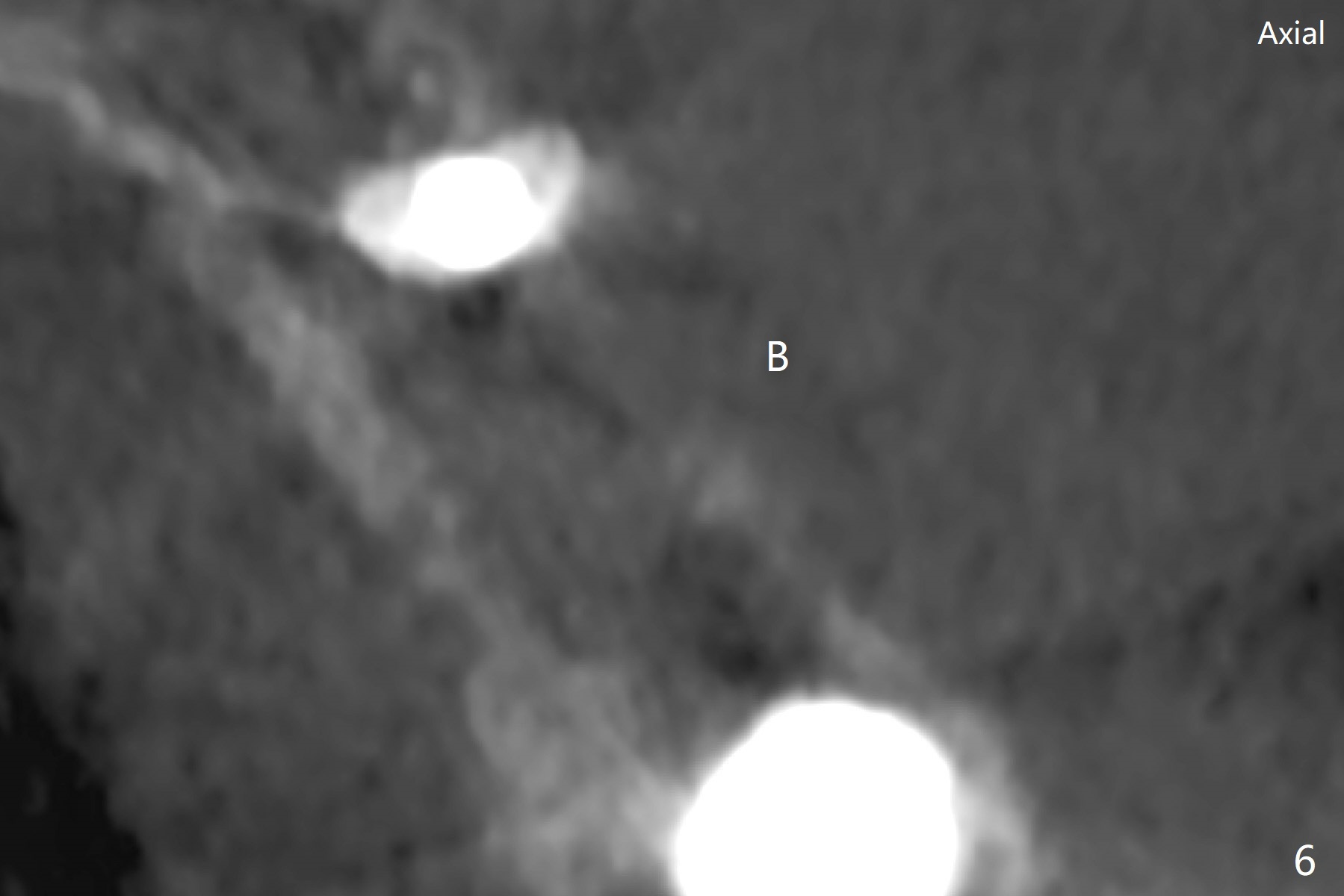
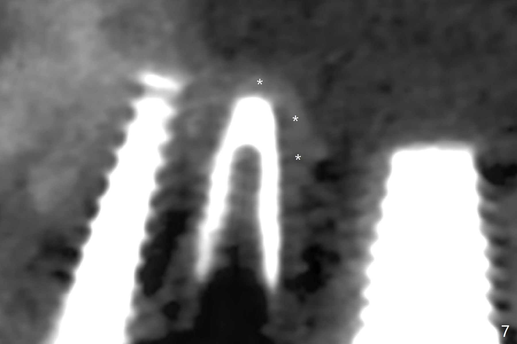
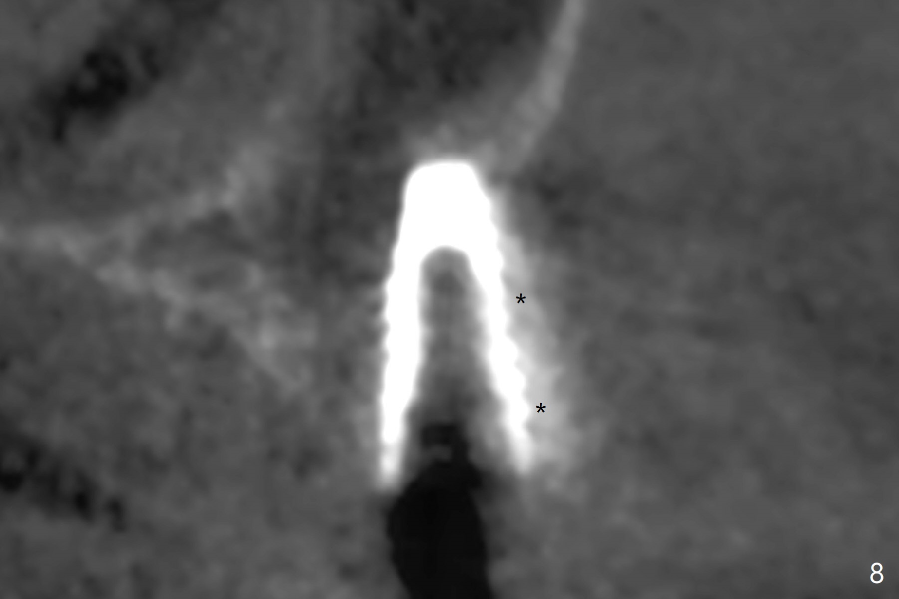
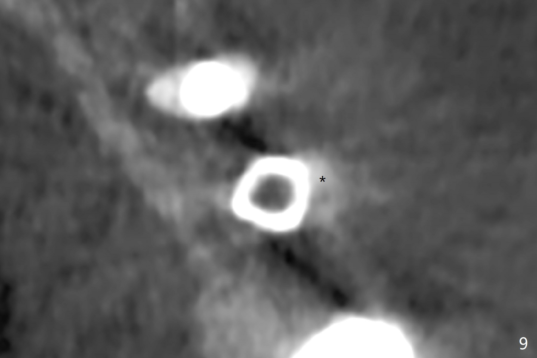
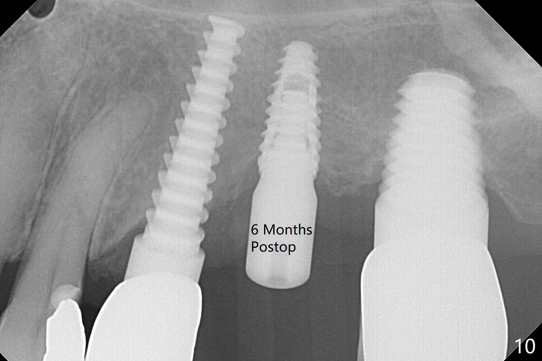
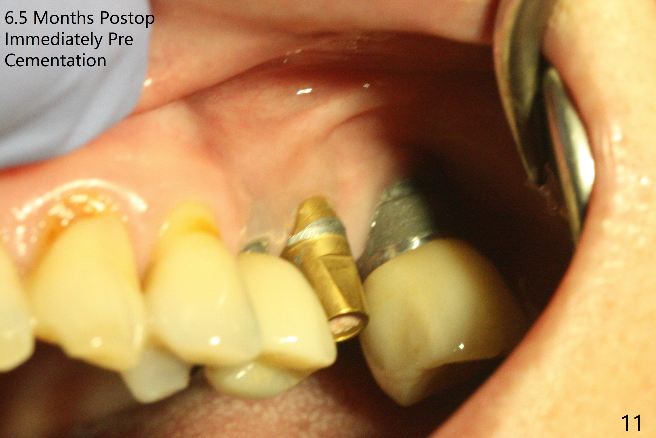
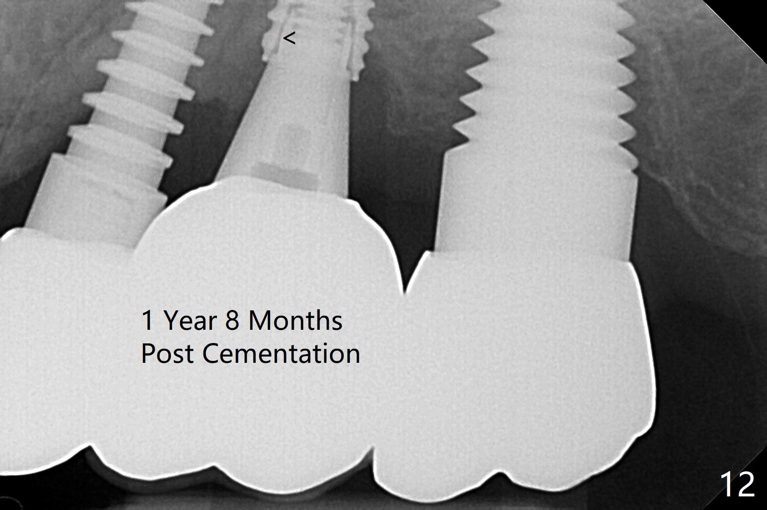
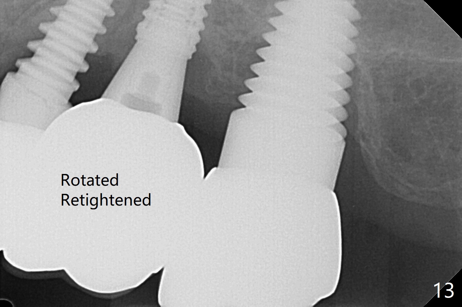
After osteotomy with guide and 2.2 mm drill for ~ 4 mm at #14, a bone expander (1.3/2.3 mm) is used free hand for sinus lift for 12 mm (from the gingival margin, Fig.1; a 10 mm long implant is planned). Following the next expander (1.7/3.1 mm) for the same depth with binding, the sinus membrane is suspected to have perforated. A piece of Osteogen Plug is inserted into the osteotomy as deep as possible with the purpose to repair the sinus membrane (no bone graft being used), followed by placing a 3.5x8.5 (instead of 10) mm implant with insertion torque ~ 25 Ncm (Fig.2,7-9, CT). As compared to preop CT (Fig.4-6), the previously grafted bone is lifted into the sinus by the bone expanders and the implant (Fig.7*). At the same time, the grafted bone has been condensed and pushed buccally (B) (Fig.8,9 *). A 4x6 mm healing abutment is inserted (Fig.3). There is crestal bone loss 6 months postop (Fig.10). Buccal plate atrophy involves the 3 implants in a row (Fig.11). The crown is loose 1 year 8 m post cementation (Fig.12). After proximal reduction, the crown/abutment rotates and sits down substantially (Fig.13). Since the crown is extremely long, it cannot be seated together with the abutment. The latter is seated with X-ray confirmation before proximal reduction of the crown (Fig.14).
Return to
Upper Molar
Immediate Implant,
Trajectory,
Next Similar Case
Torque 20 Ncm
2Xin Wei, DDS, PhD, MS 1st edition
01/22/2019, last revision
04/30/2021