|
|
|
|
|
|
|
|
|
|
|
|
|
|
|
|
|
|
|
|
|
|
|
|
|
|
Ball Abutments for Maxilla
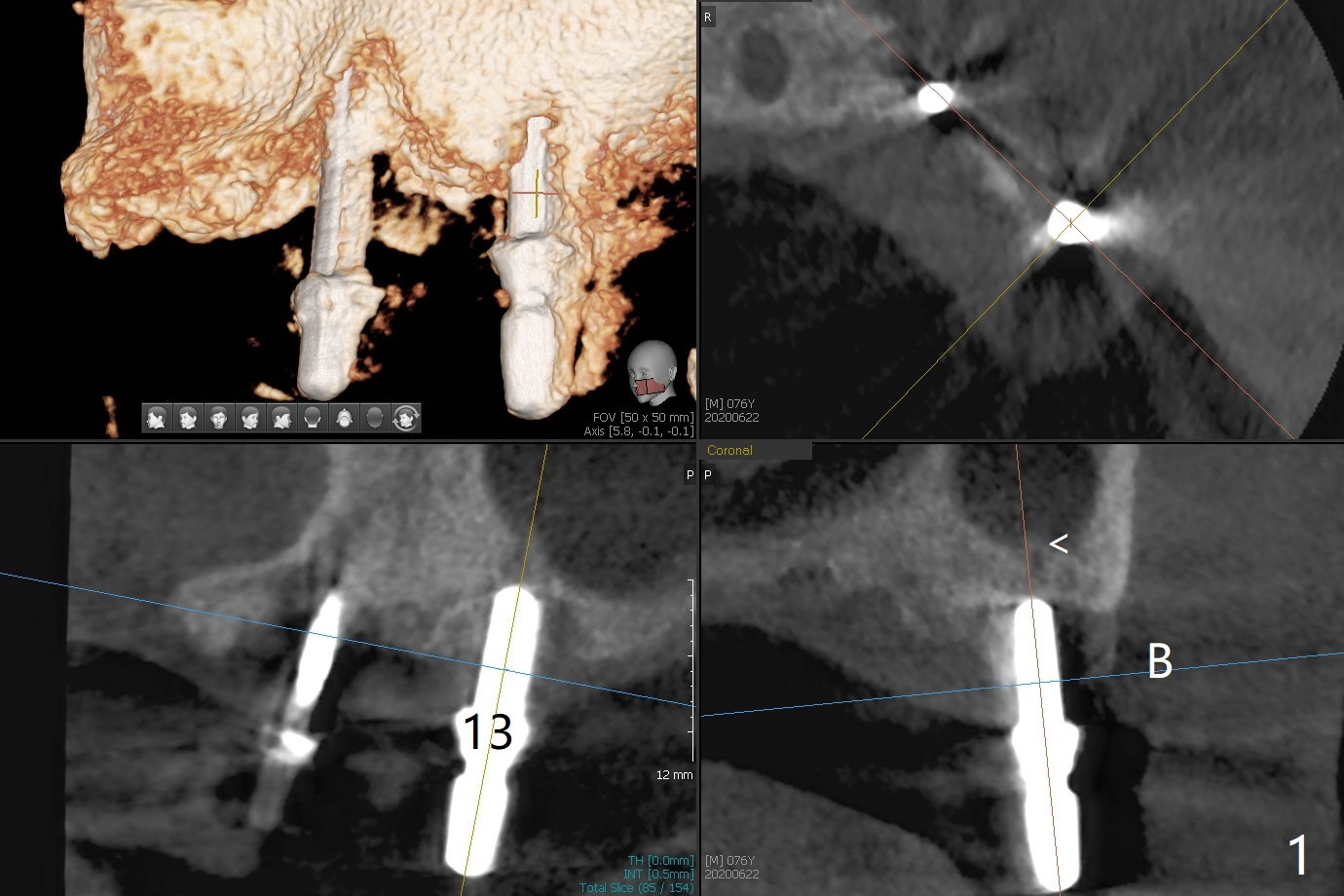
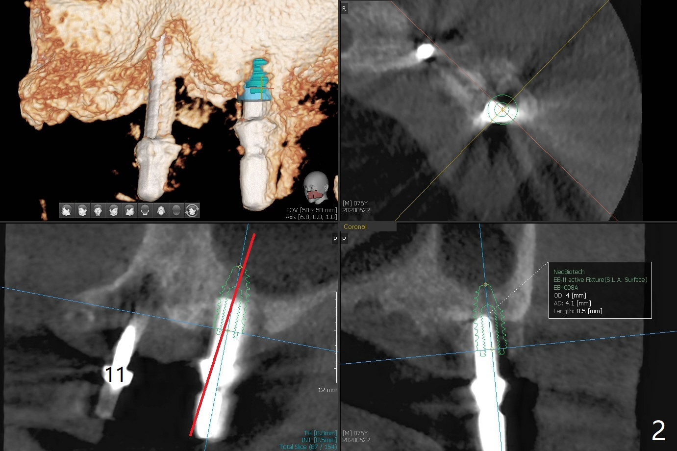
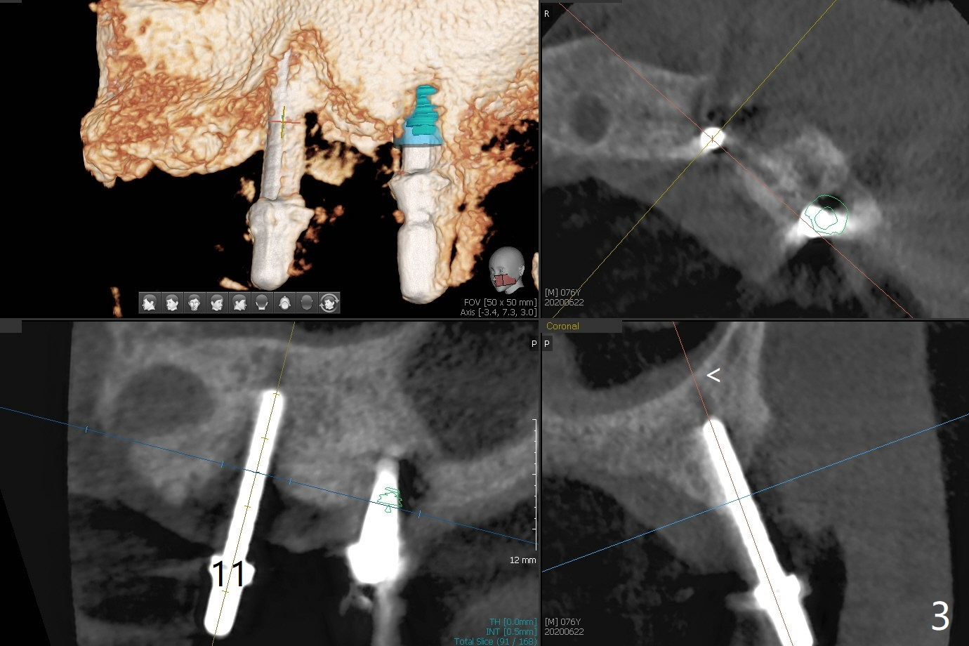
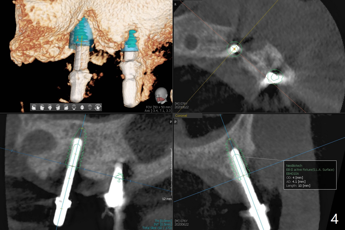
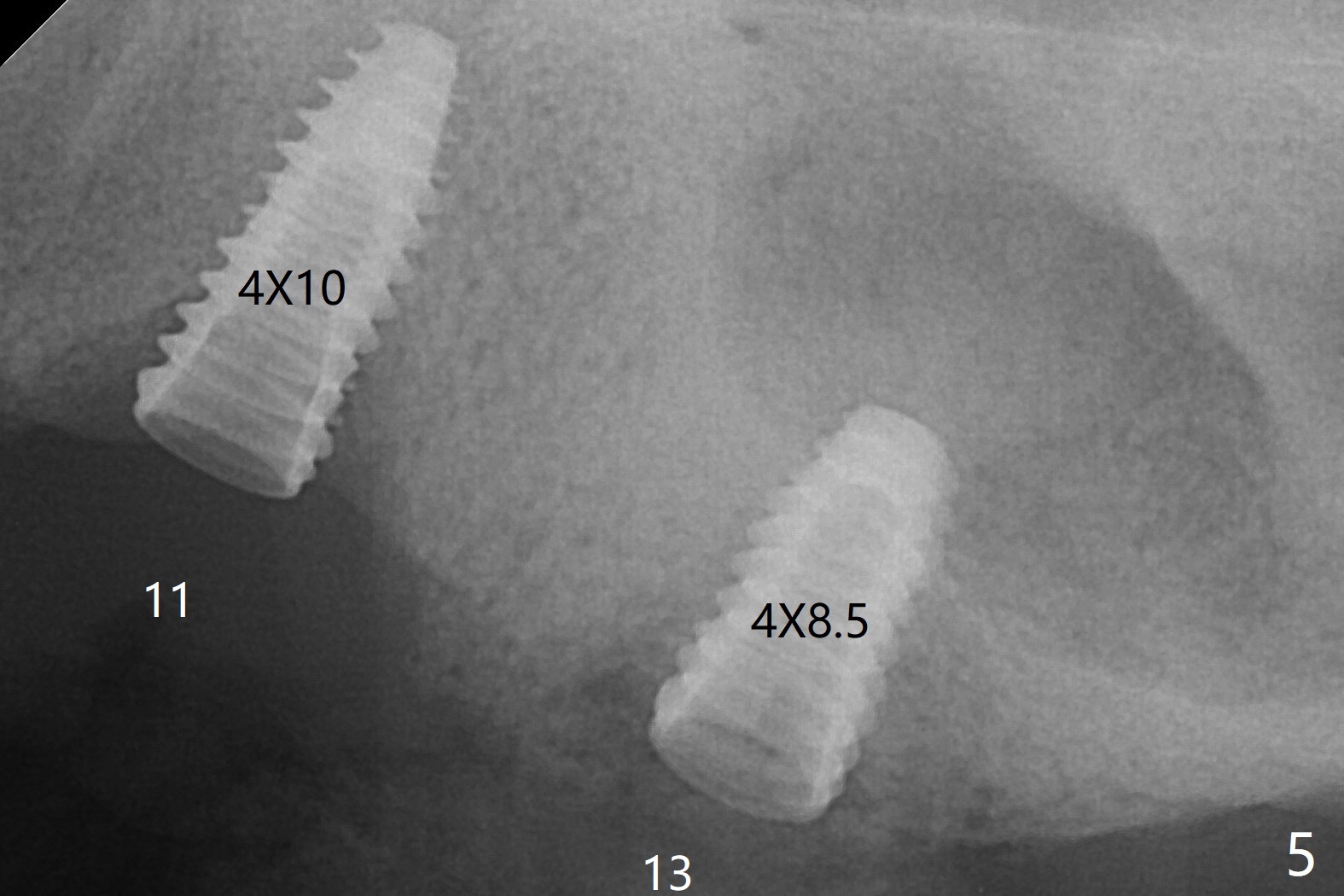
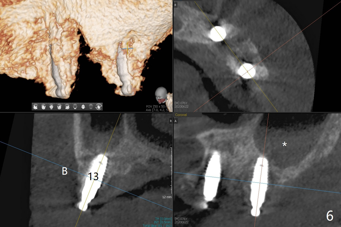
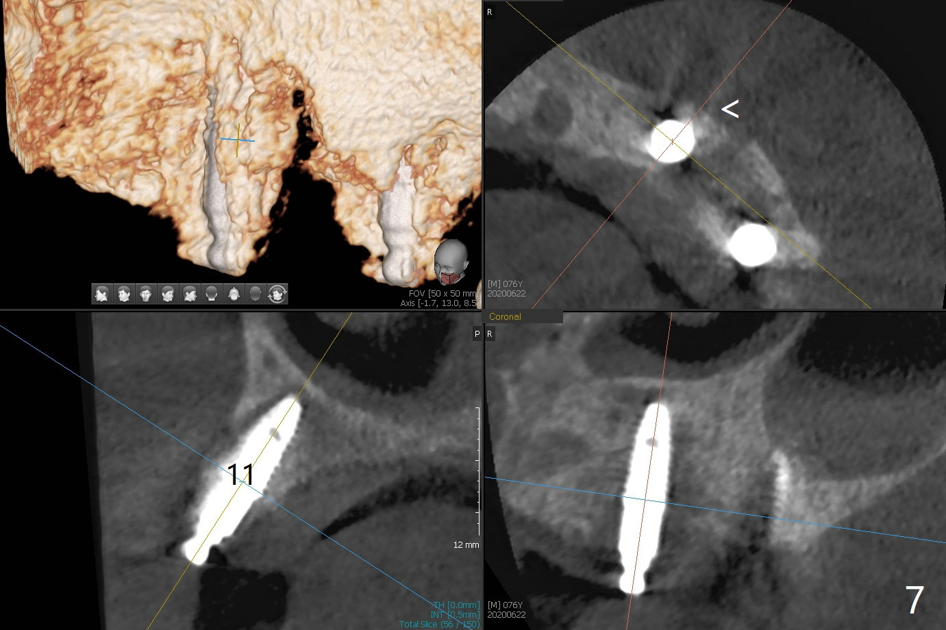
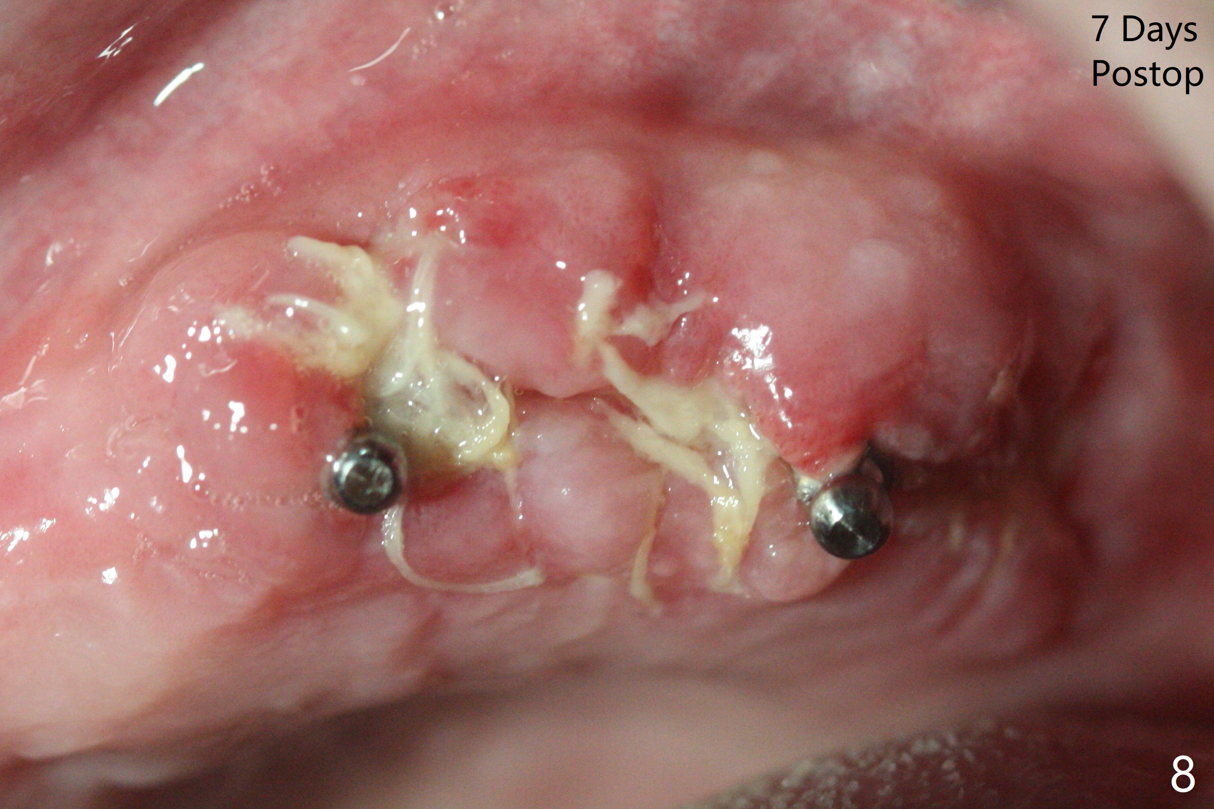
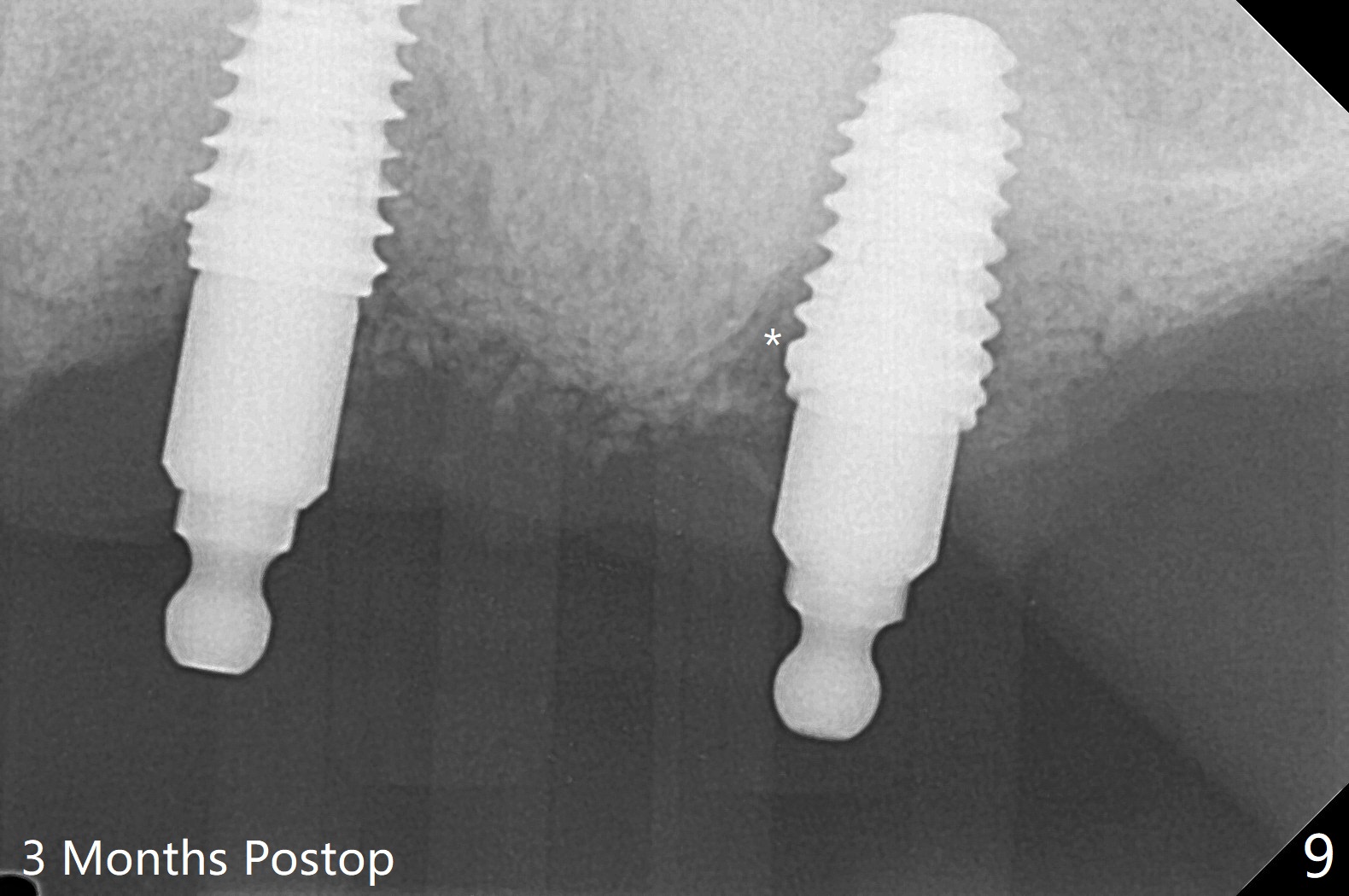
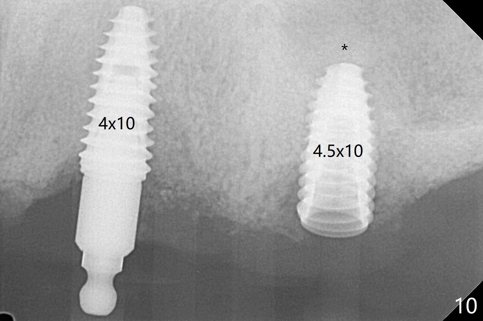
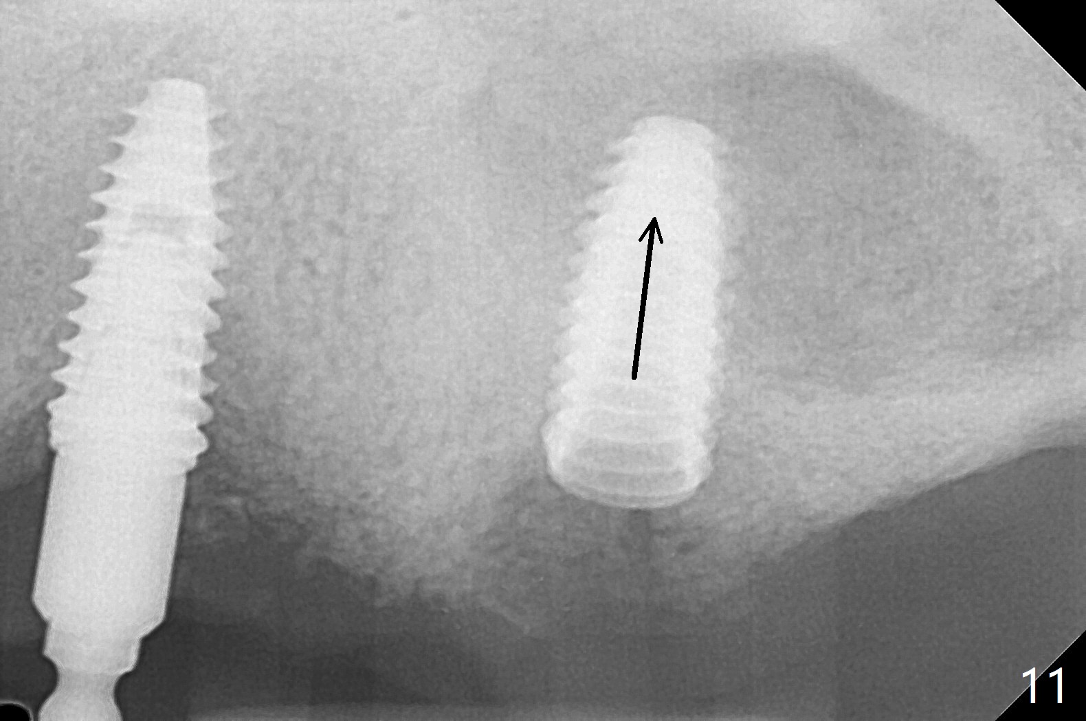
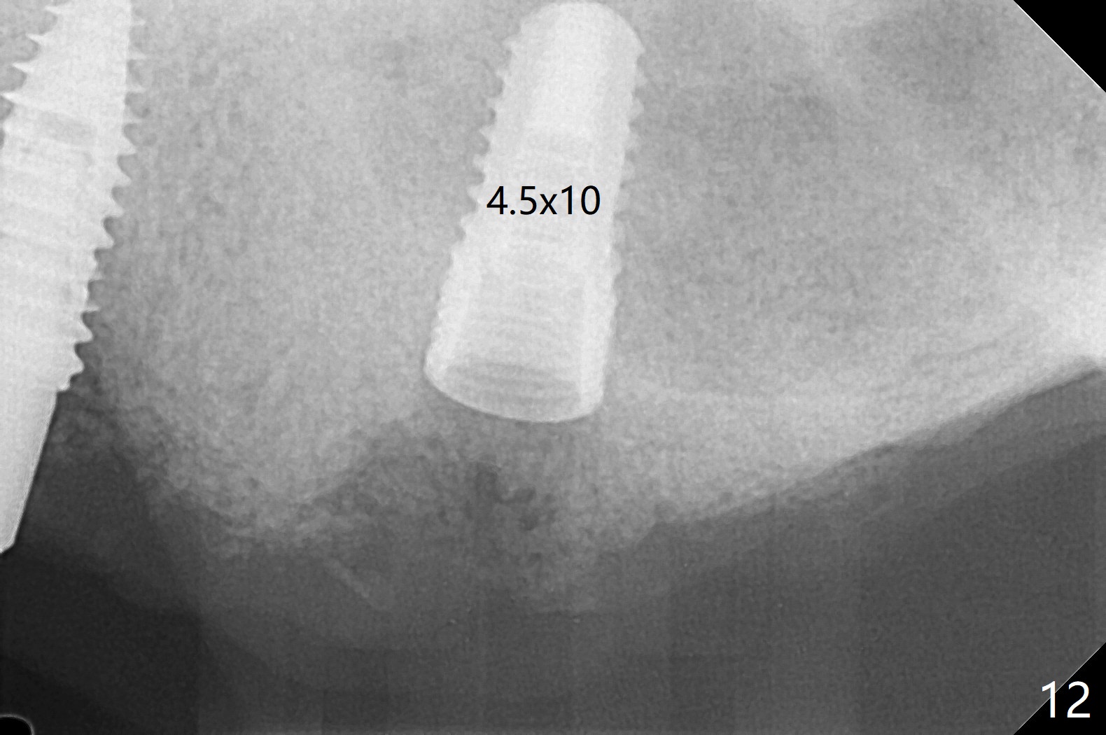
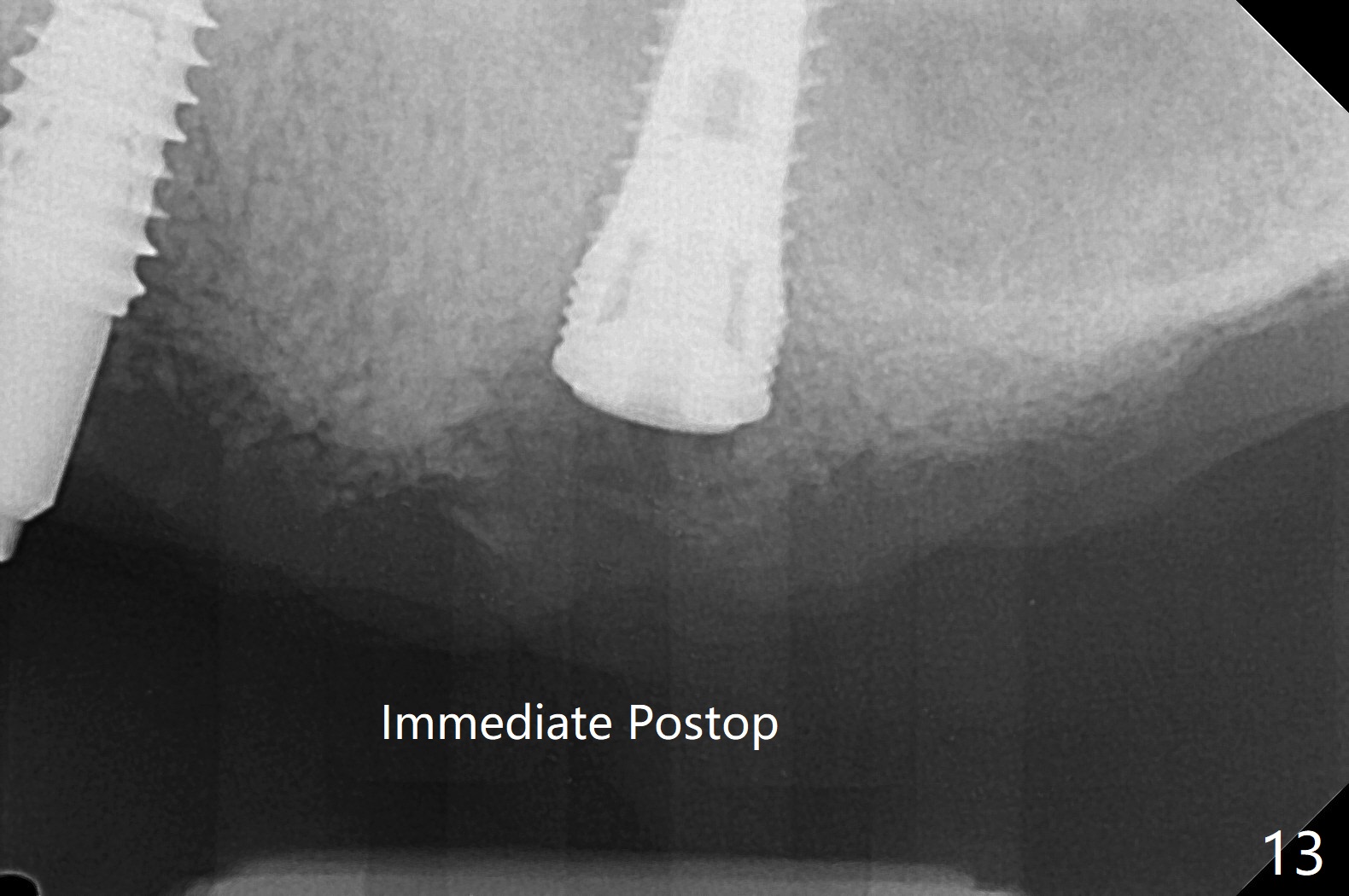
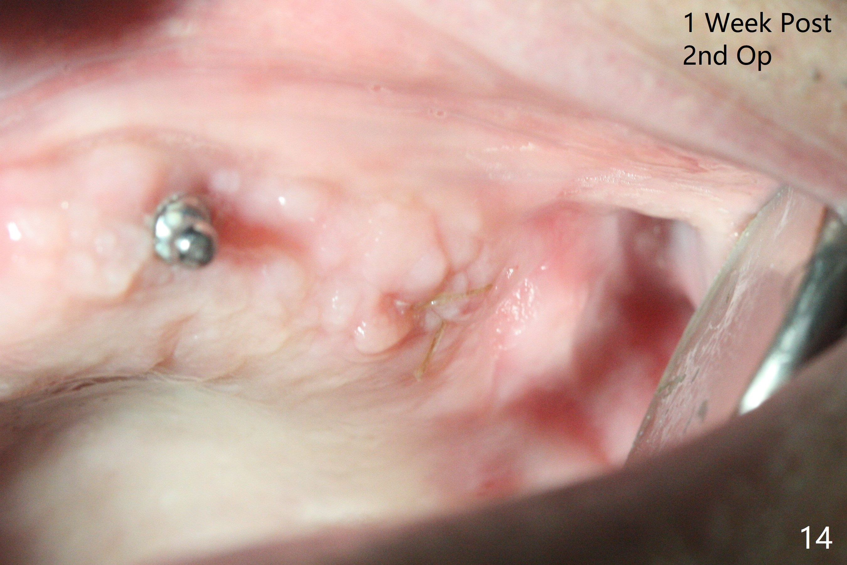
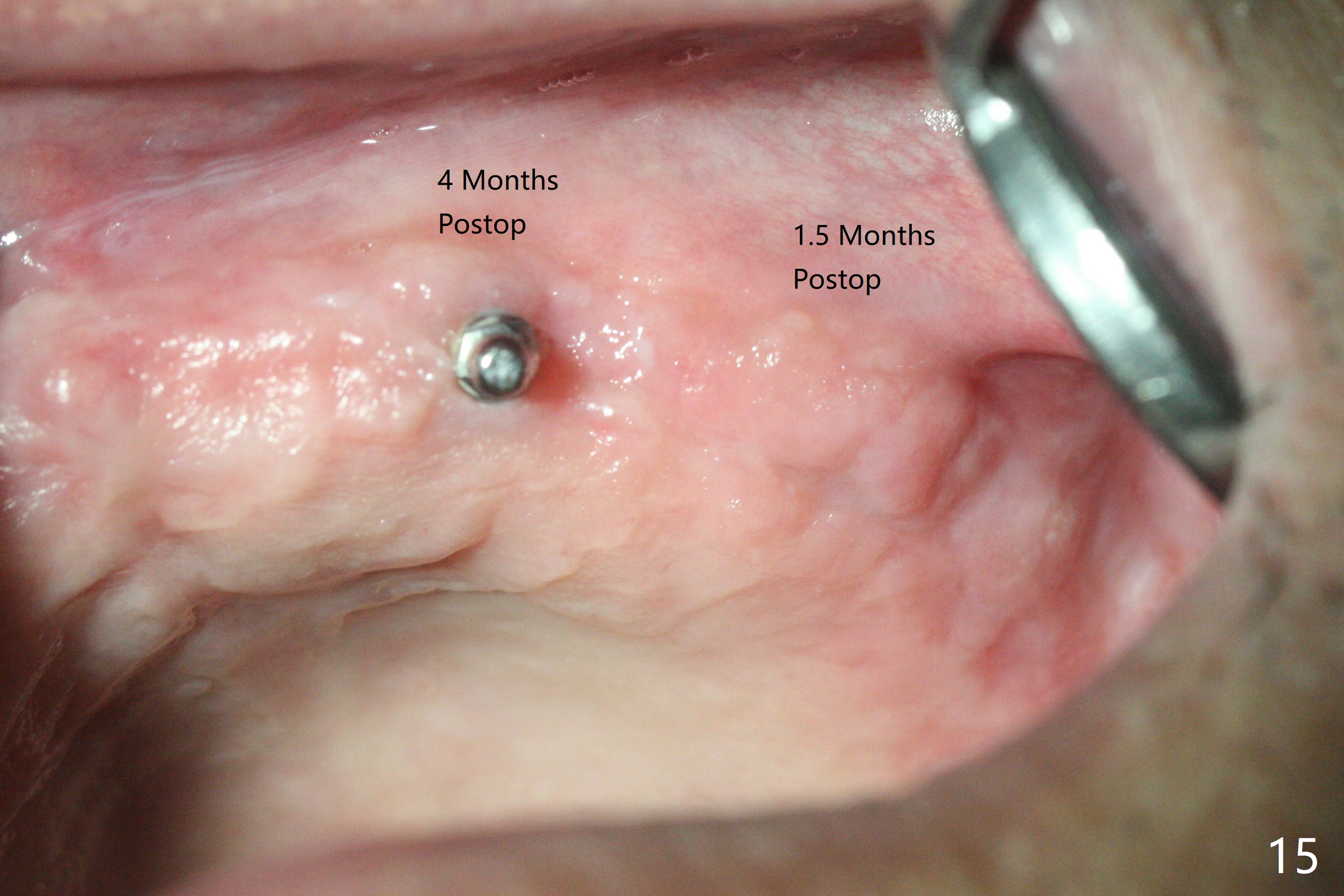
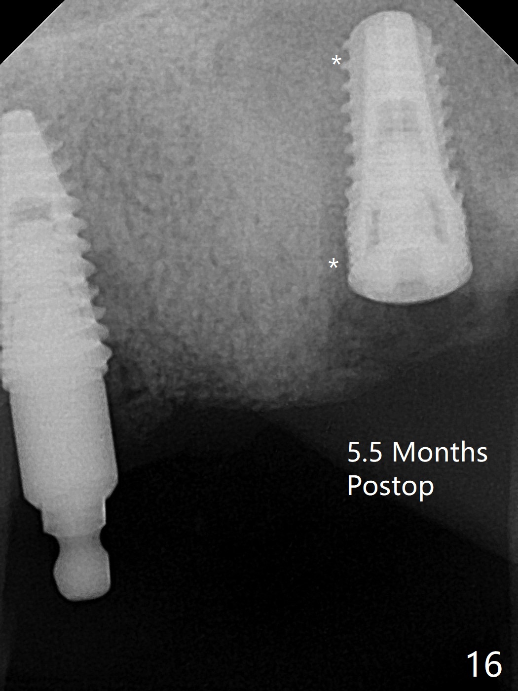
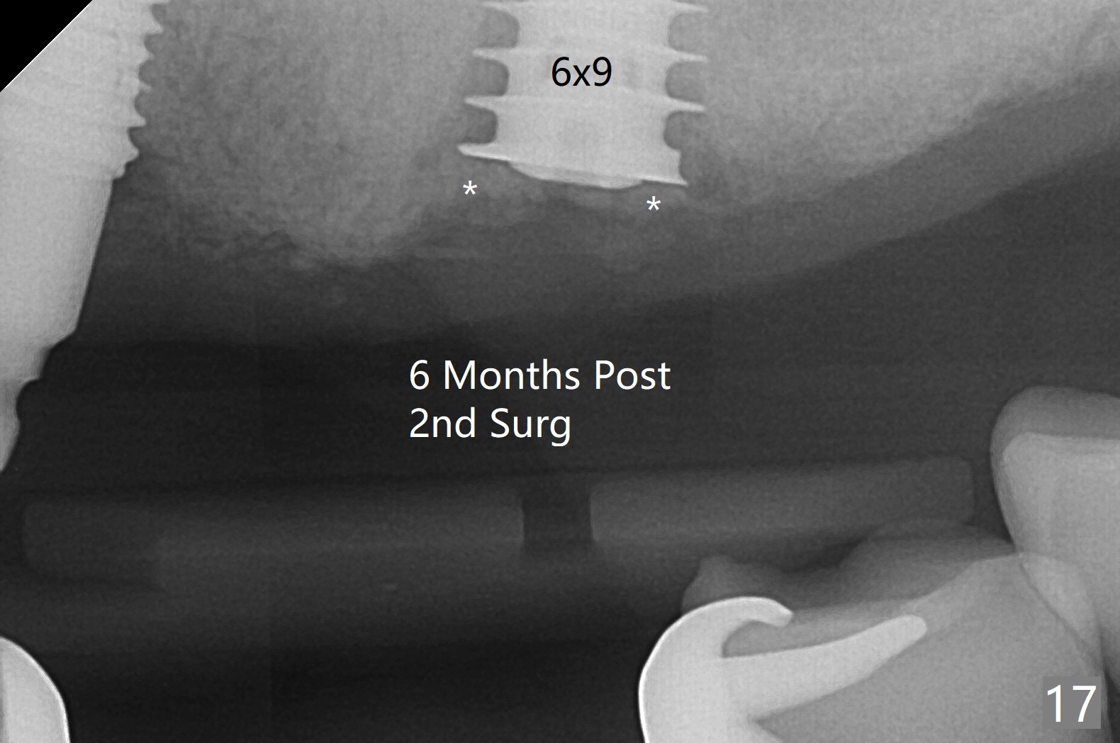
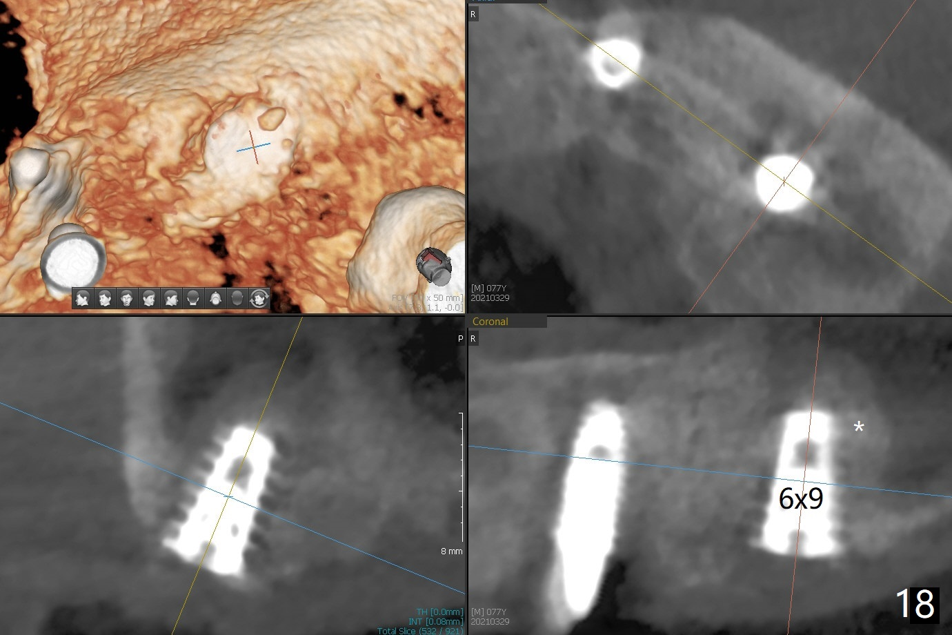
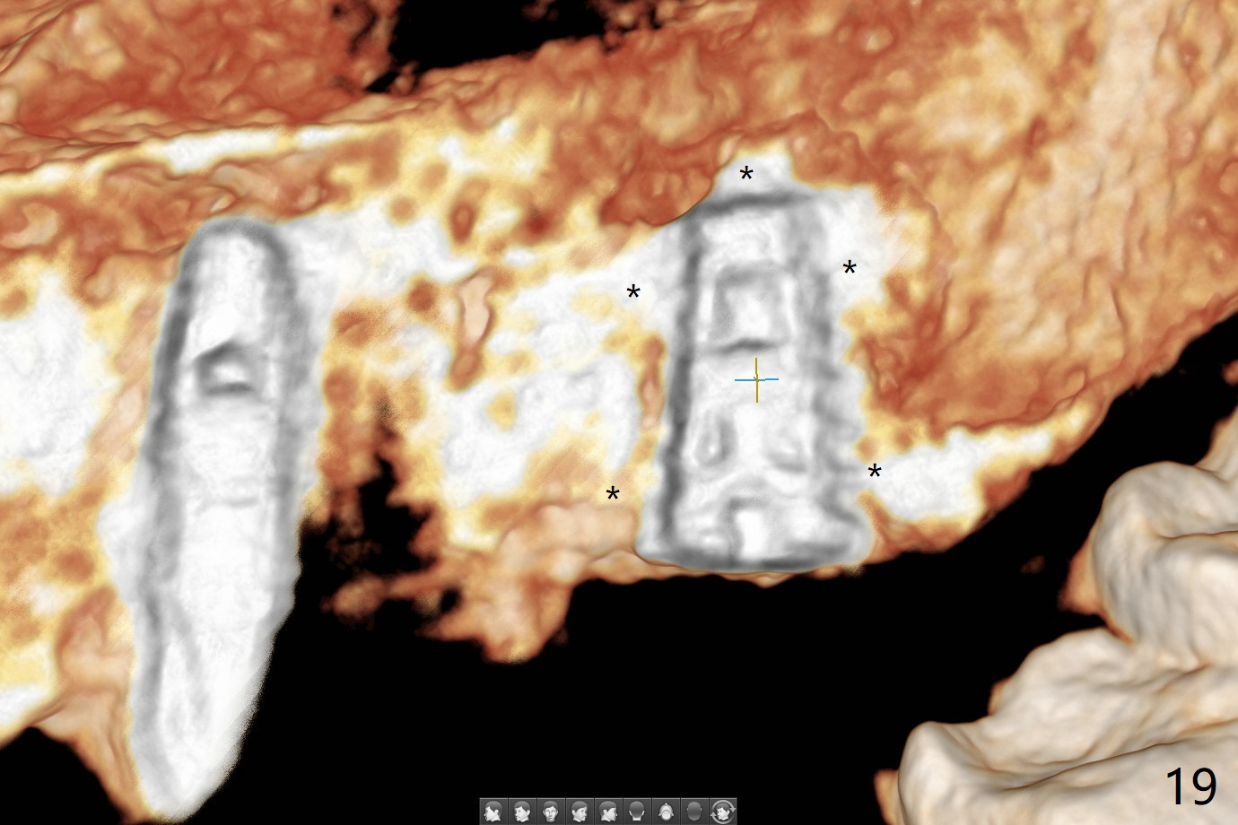
Osteotomy at #13 starts blindly (no incision or tissue punch) with bone expanders. After insertion of parallel pins, intraop CT shows that it is palatal with apparently buccal low bone density (Fig.1); the osteotomy for implant (Fig.2 green) should be shifted buccal and tilted mesial (red, parallel to #11) with incision. In contrast the position, trajectory and depth (Fig.3 <) of the initial osteotomy are acceptable at #11. The implant will be placed as it is (Fig.4). After taking a postop PA (Fig.5), the implant at #11 is placed a little deeper to make sure its slightly subcrestal placement (including distal incision at #11). Following placement of 3.5x4 and 3 mm ball abutments at #11 and 13, cortical allograft with PRF is placed around the implants, especially buccal (Fig.6,7 <). After suturing, the profile of the ball abutments is too low for RPD retention. Due to gravity other than bone density, the number of ball abutments for the maxilla should be more than for the mandible. Soft reline is done to the patient's satisfaction. The retention of the upper RPD after soft reline is satisfactory without pain 7 days postop (Fig.8). The implant at #13 is loose nearly 3 months postop (Fig.9 *: bone loss). The implant is removed while the ball abutment is untightened; the sinus floor is present. It appears that a longer and larger implant is necessary; a 4.5x10 mm dummy implant is unable to be seated deep or achieve primary stability (Fig.10). After sinus lift with 3 mm Bicon osteotome without bone graft, the dummy implant accomplishes the 2 tasks mentioned above (Fig.11). However there is no corresponding definitive implant in stock. Implant system needs to be changed; with a change in implant driver, the depth control is lost. The final implant is placed deep (Fig.12). With back up, stability is lessened; a healing screw is placed; with collagen plug, the wound is sutured (Fig.13). The wound heals 1 week postop (Fig.15). The RPD is soft relined. Retention from the ball abutment at #11 is apparently critical. The RPD and #11 implants (4 months postop) are stable, while the wound at #13 heals 1.5 months postop (Fig.15). There is space around the implant 5.5 months postop (Fig.16 *). The 5x10mm SM implant is found to be loose upon uncover and removed. After debridement, 5.3x8 mm SM and 5.5x9 mm IBS dummy implants are inserted without stability, while 6x9 mm definitive one with stability (Fig.17). Cortical allograft is placed in deficiency areas (*). The osteotomy has no roof (sinus floor), but the sinus membrane is intact. Small amount of bone graft (Fig.18 *) is placed before implantation. There appears to be bone around the new implant (Fig.19 (3D sagittal section) *).
Return to No Deviation Overdentures 劈开术 Xin Wei, DDS, PhD, MS 1st edition 06/22/2020, last revision 04/05/2021