,%203.8x18,%204.5x7(5).jpg)
,%203.8x18,%204.5x7(5).jpg)
|
|
|
|
|
|
|
|
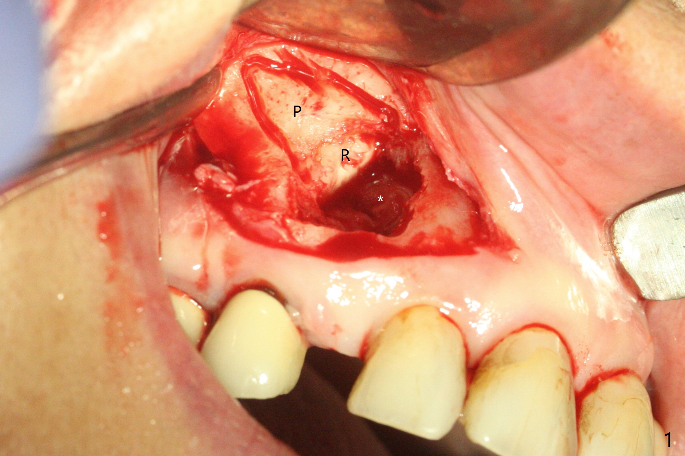
Difficult to Extract Impacted Canine
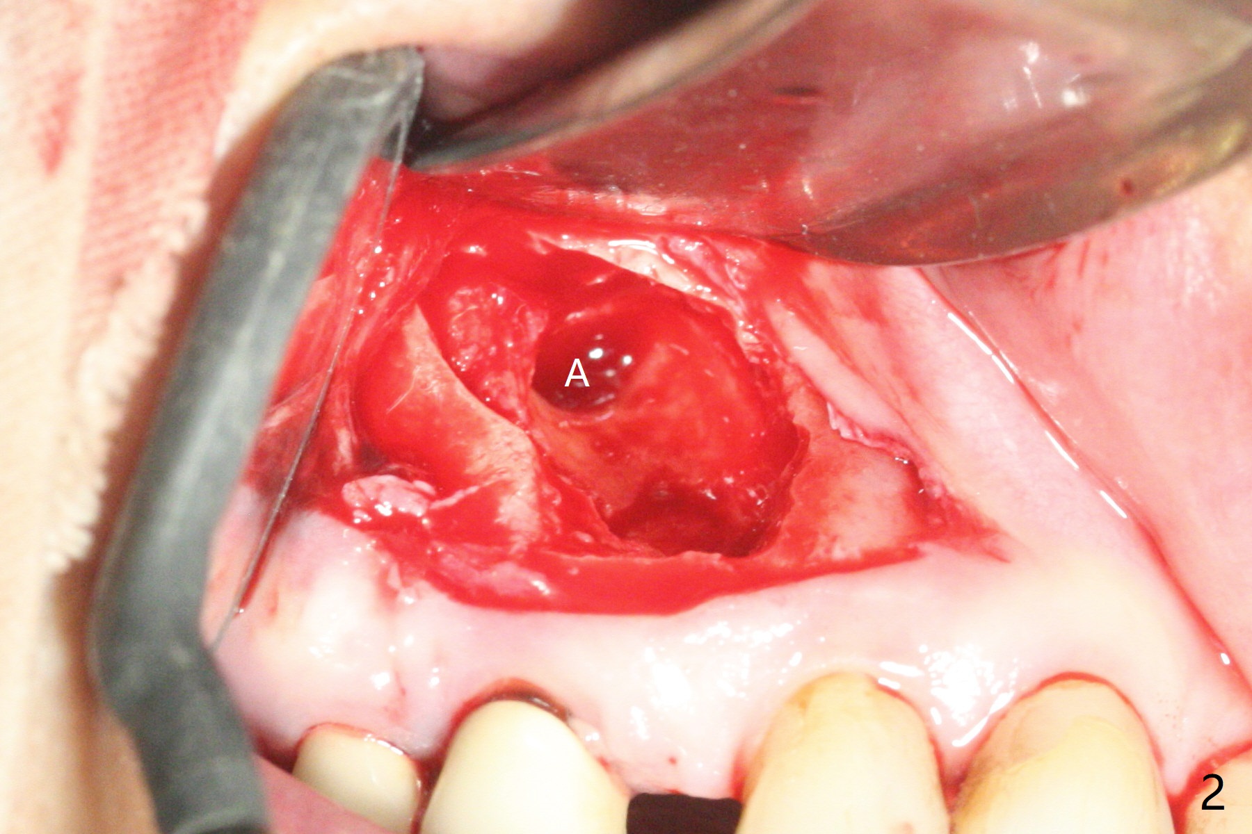
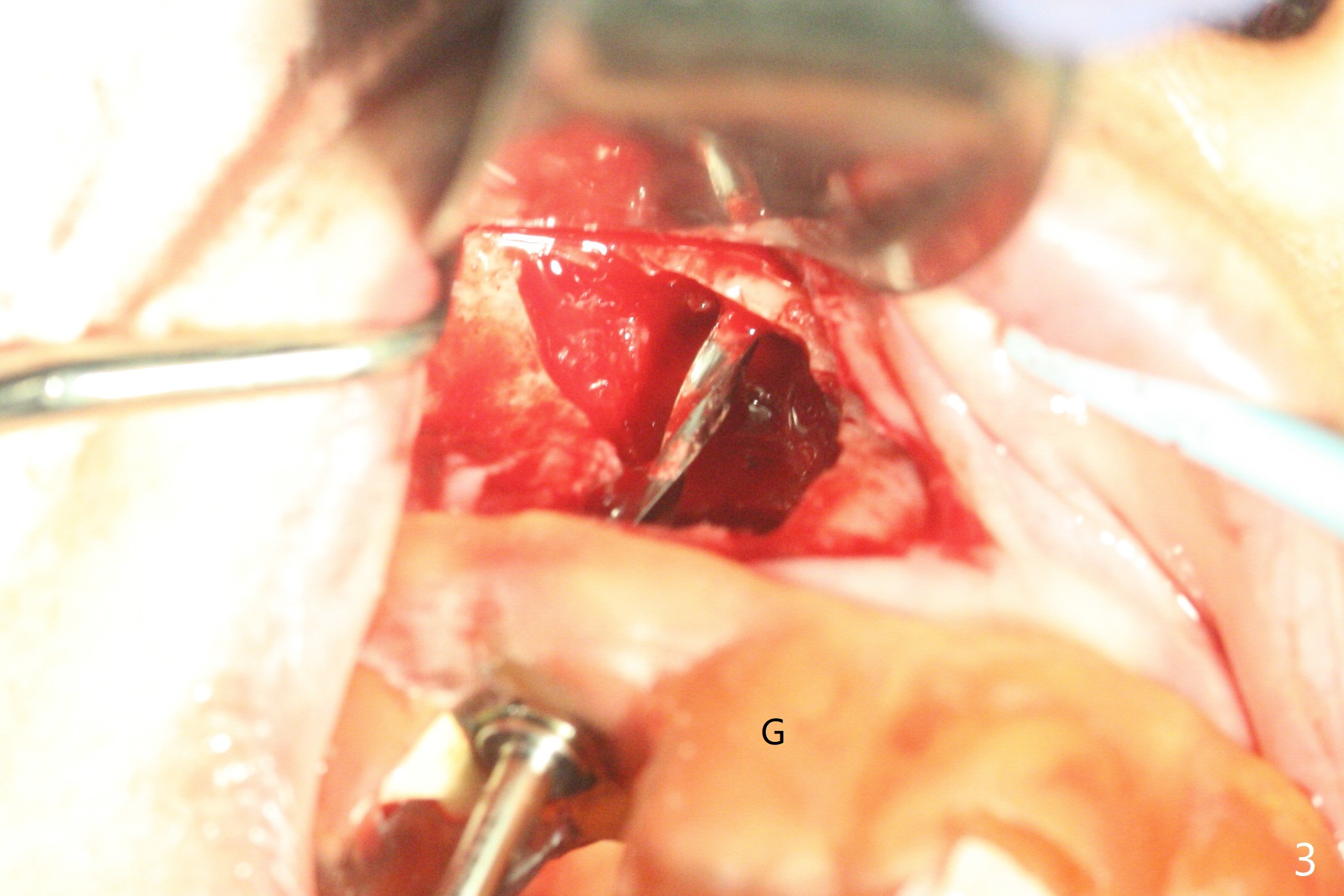
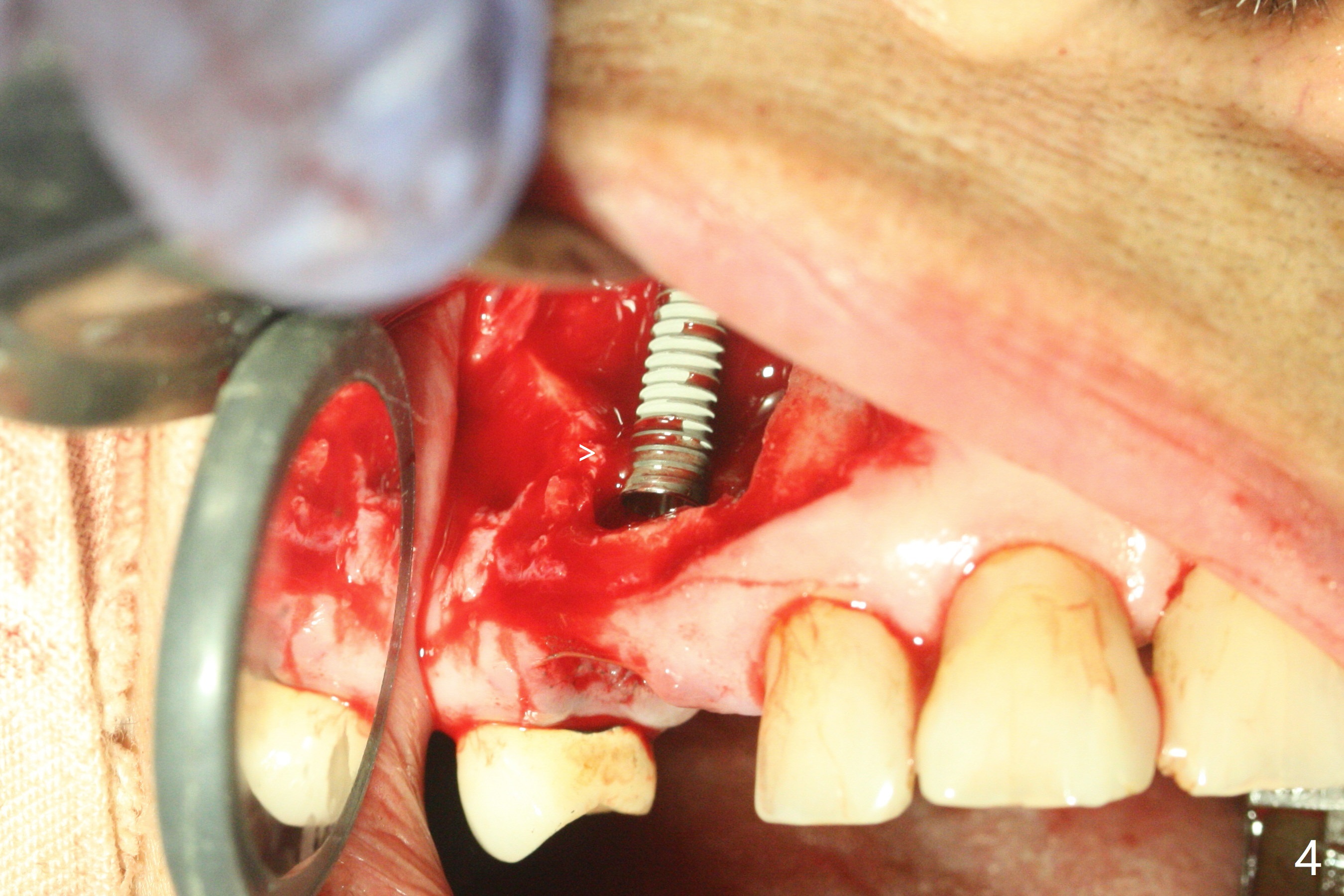
After incision and flap elevation, the crown of the impacted canine is more superficial. With removal of the buccal cortical plate and multiple sectioning of the crown, the latter is removed (Fig.1 *). Further removal of the buccal plate (P) and purchase points, the root (R) is extracted easier. In fact the apex of the root is the deepest (Fig.2). With the guide (Fig.3 G) and 2.2 mm drills, the osteotomy is established. It is pretty shallow. A 3x18 mm drill is used to deepen the osteotomy free hand (mistake: too palatal). When a 3.8x18 mm UF implant is being placed, it cannot be placed deep with the guide. When the latter is removed, the implant seems to be placed too deep and too buccal with <10 Ncm (Fig.4). A 4.5x7(5) mm abutment is placed mainly to correct the trajectory of the implant to certain degree. Placement of the implant at #3 is quite smooth (Fig.5) in spite of severe bone loss and abundant granulation tissue at #3 and 5. The implant at #5 does achieve 2 pointed fixation (Fig.4,6 arrowheads). The most coronal portion of the canine crown is left behind (Fig.6 C).
Return to
Prevent Molar Periimplantitis (Protocols,
Table)
Protect
Graft
Torque
Oral
Scanner 智齿拔除
Xin Wei,
DDS, PhD, MS 1st edition 06/24/2021, last revision
07/25/2021