.jpg)

|
|
|
|
|
|
|
|
|
|
||
|
|
|
|
|
|
|
|
|
|||
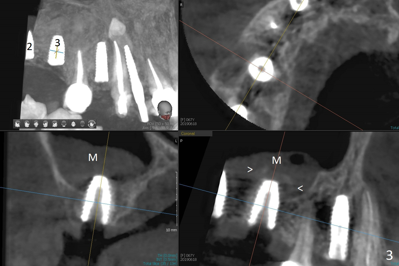
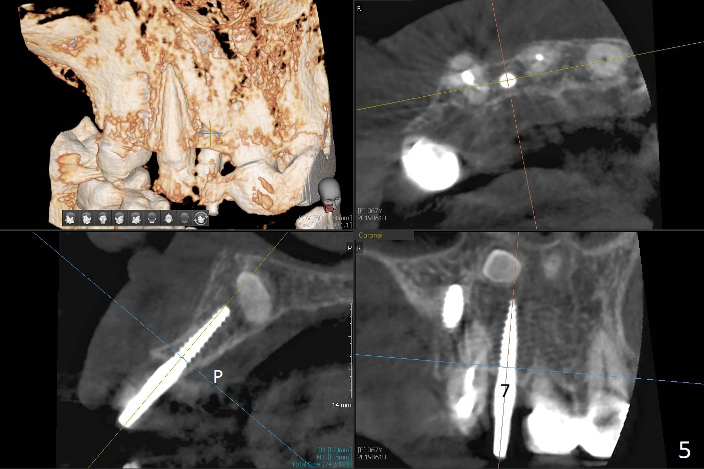
Swollen Sinus Membrane
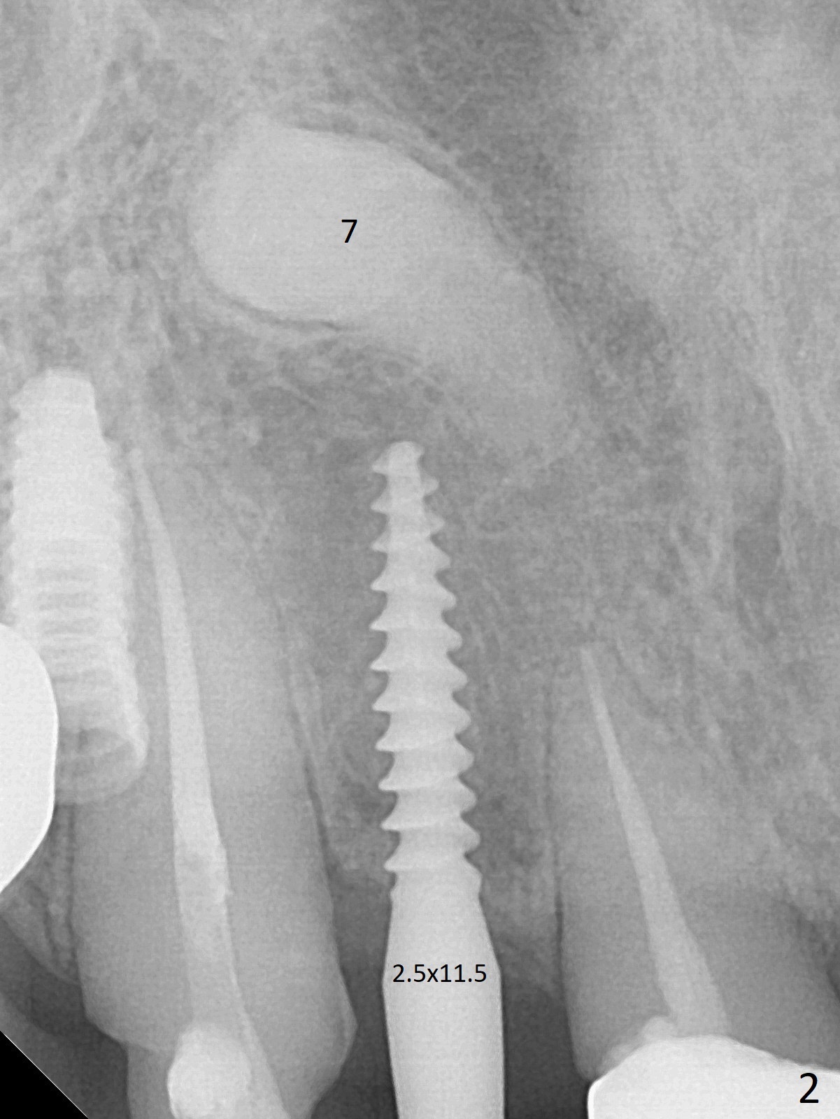
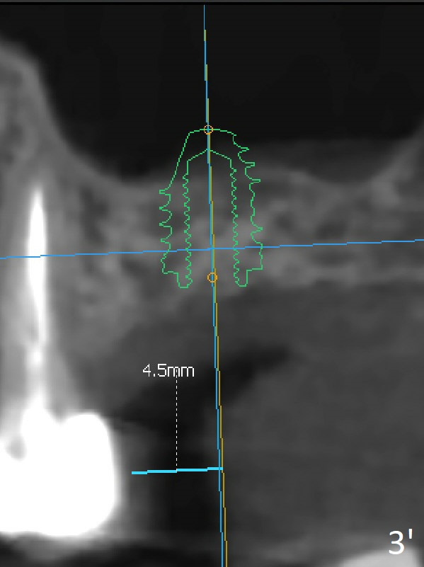
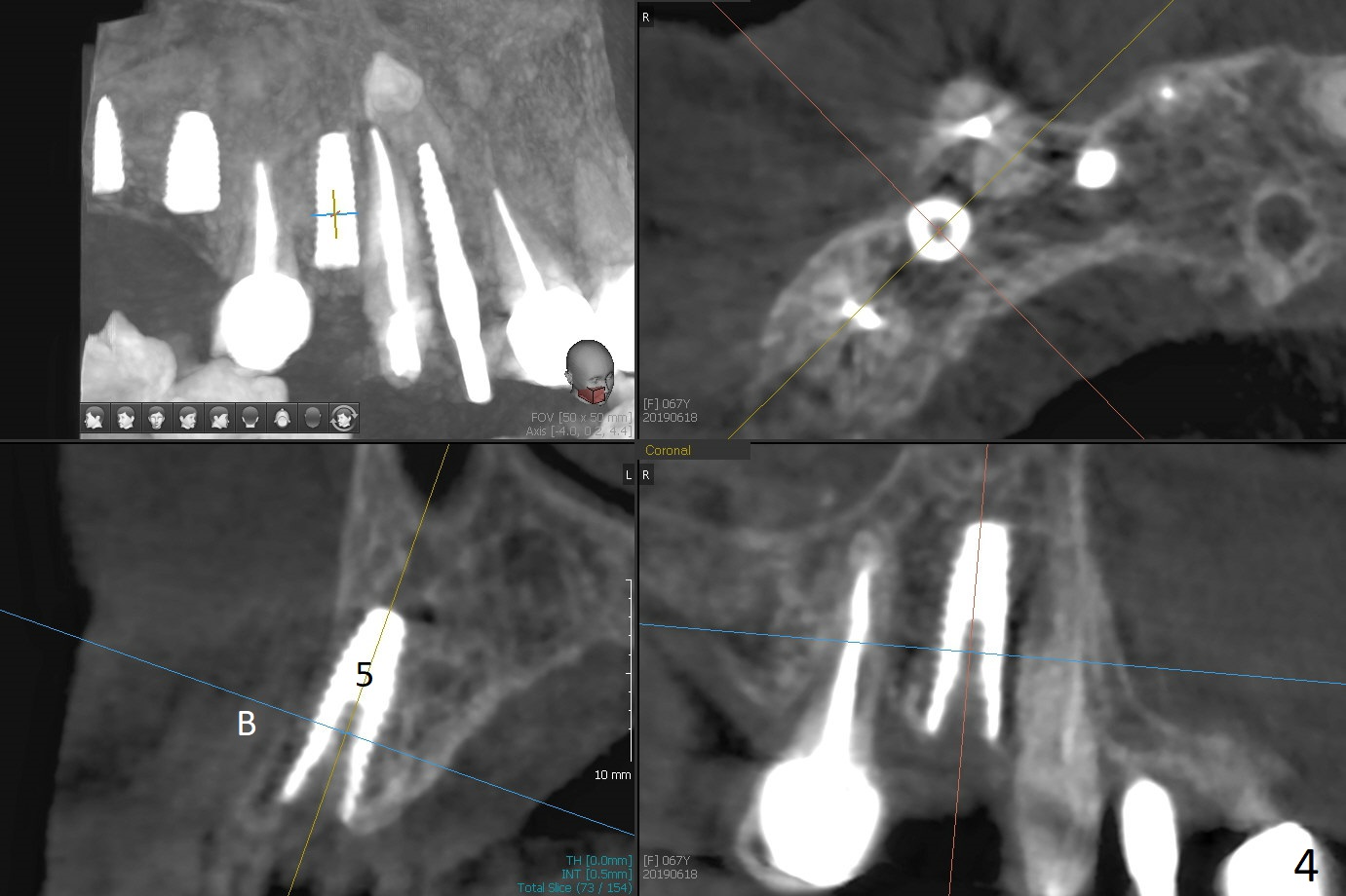
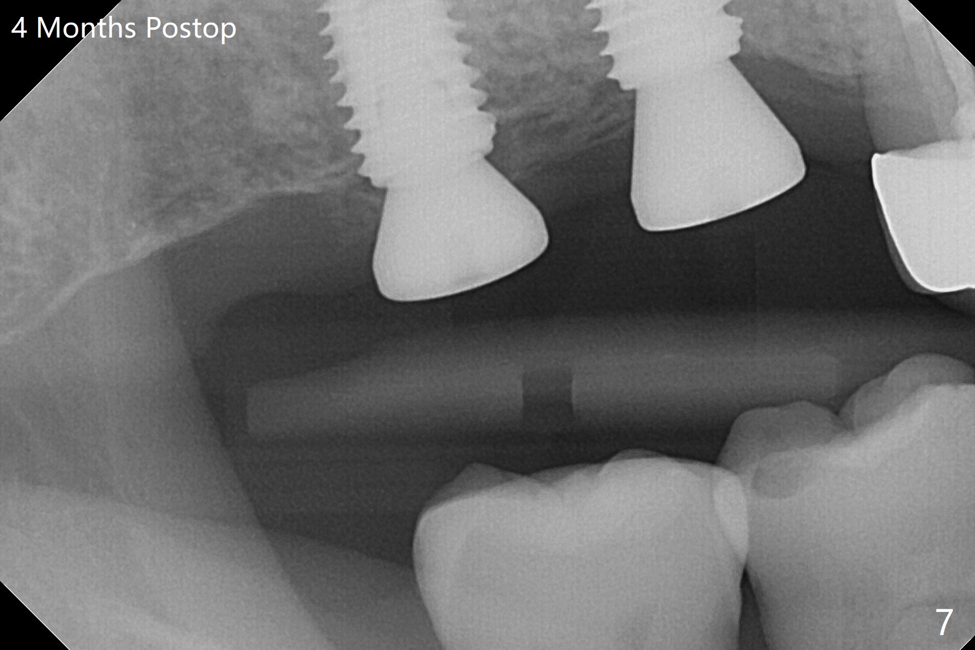
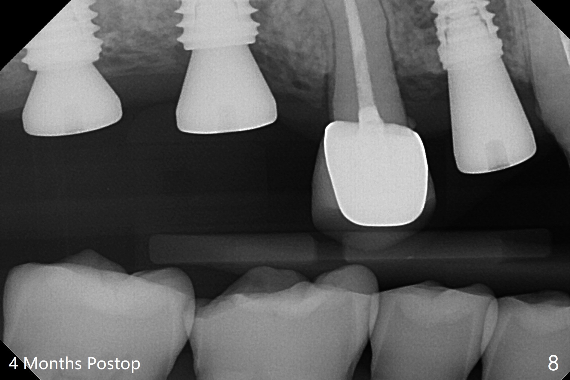
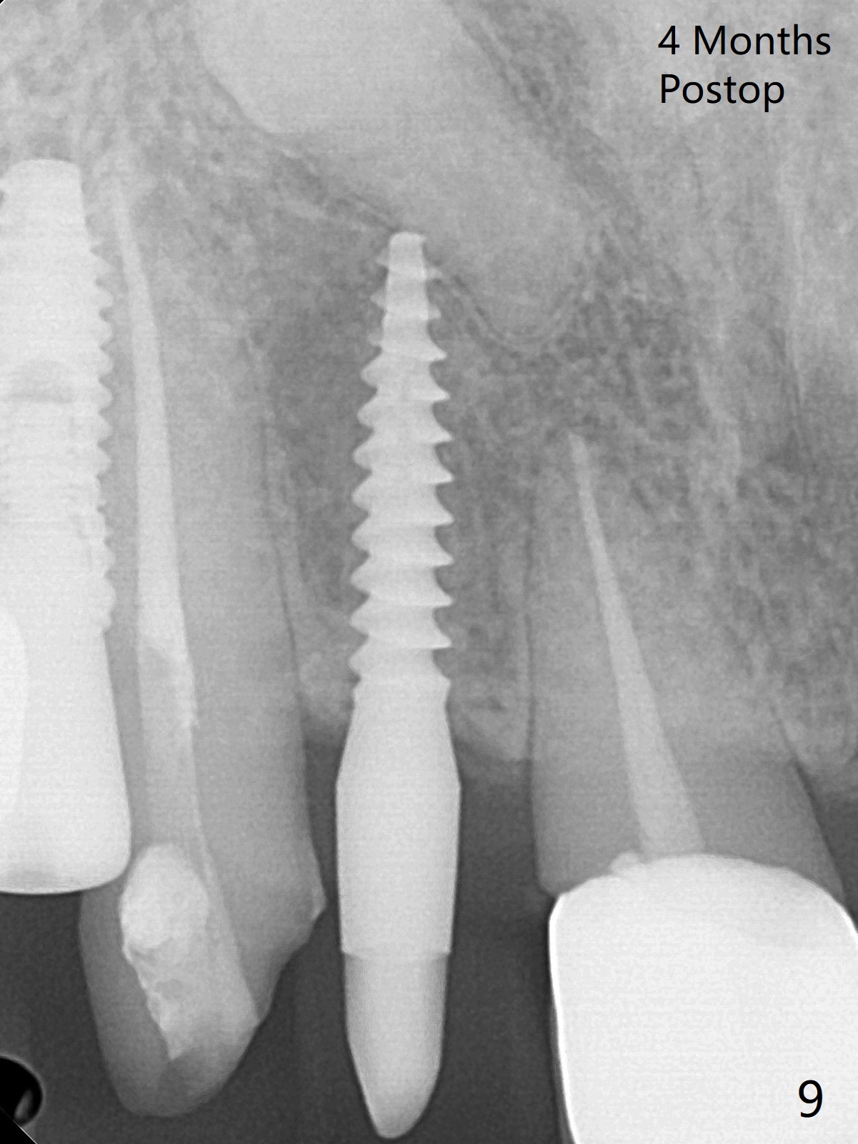
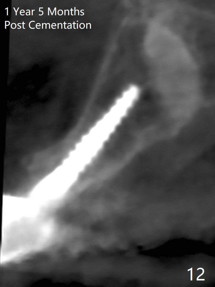
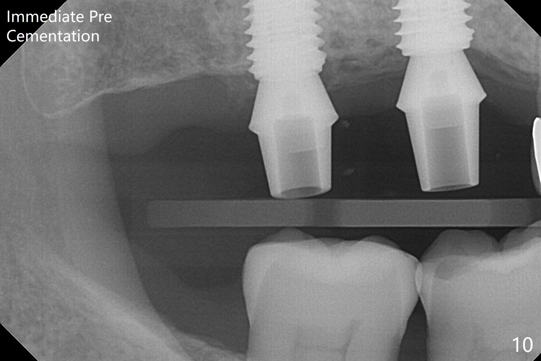
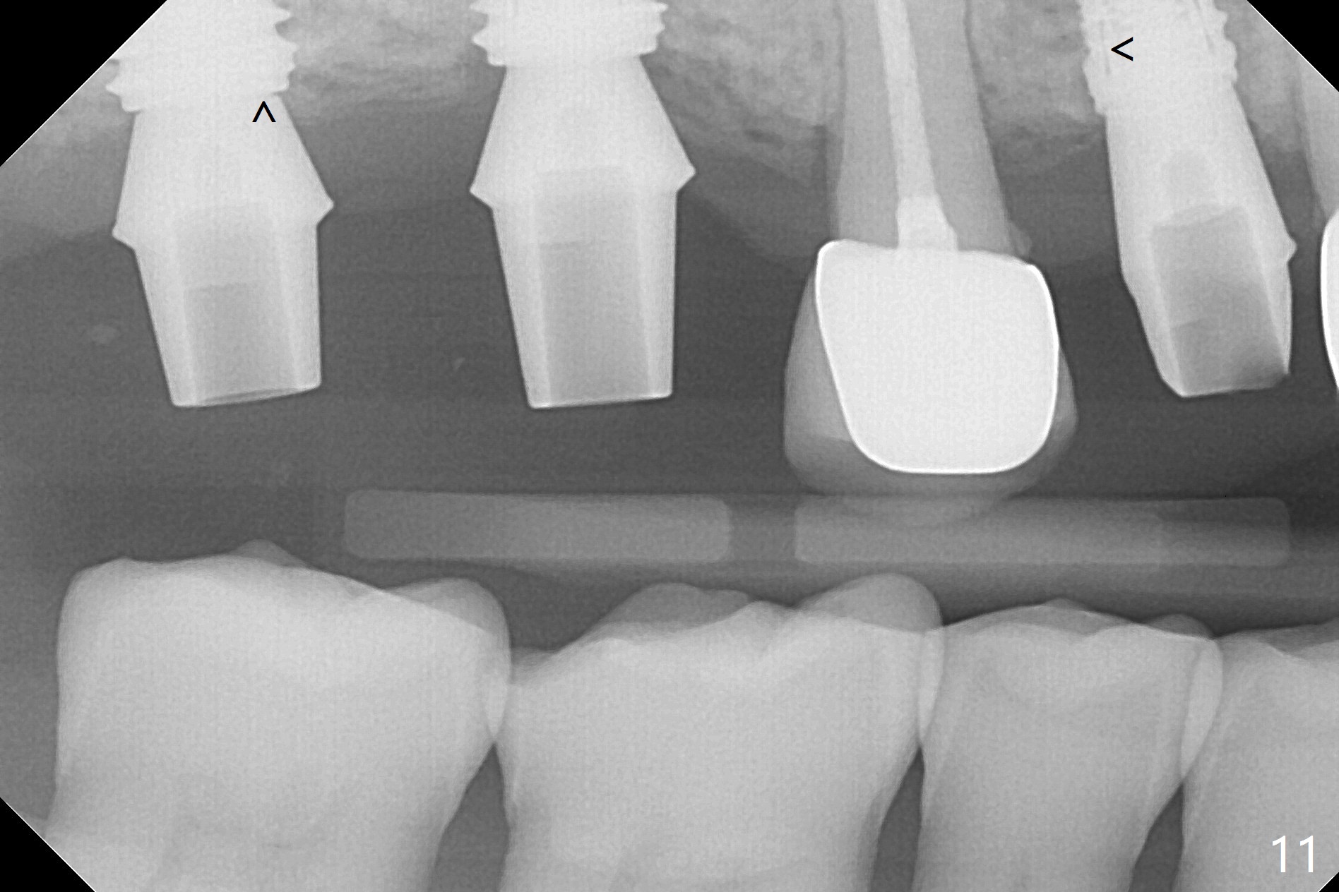
Two of 5x8.5 mm implants are placed at #2 and 3 with sinus lift using water ballooning technique; to avoid invading the neighboring root tips, an intraop PA is taken with a 2.2x11.5 mm drill at #5 (Fig.1). When a 2.5x11.5 mm 1-piece implant is placed at #7, there is 2.4 mm clearance from the impacted tooth (Fig.2). Following 1 mm deeper placement of the 1-piece implant, CT is taken to make sure that the threads are completely covered palatally (Fig.5 P). Interesting is that the sinus membrane is thickened at #2 and 3 (Fig.3 M), as compared to the clear sinus before surgery (Fig.3'). Bone graft is limited in the sinus (Fig.3 arrowheads). The implant at #5 (3.5x11.5 mm) is precisely placed subcrest bucco(B)-palatally (Fig.4). Since the limited field viewed CT does not cover the implant at #2 (Fig.3), immediate postop PA is taken (Fig.6). In fact all of the implants are placed subcrestal, as shown by intraoral X-ray 4 months postop (Fig.7-9). The abutments at #2, 3 and 5 are retorqued (30 Ncm) after crowns' try in and adjustment (Fig.10,11). There is a vertical gap between the implant and abutment at #2 (Fig.11 ^) and a horizontal one at #5 (<). There is apparently no bone loss 1 year 5 months post cementation (Fig.12).
Return to
Full Mouth Immediate Implant,
Trajectory
12
Xin Wei, DDS, PhD, MS 1st edition
06/18/2019, last revision
04/15/2021