|
|
|
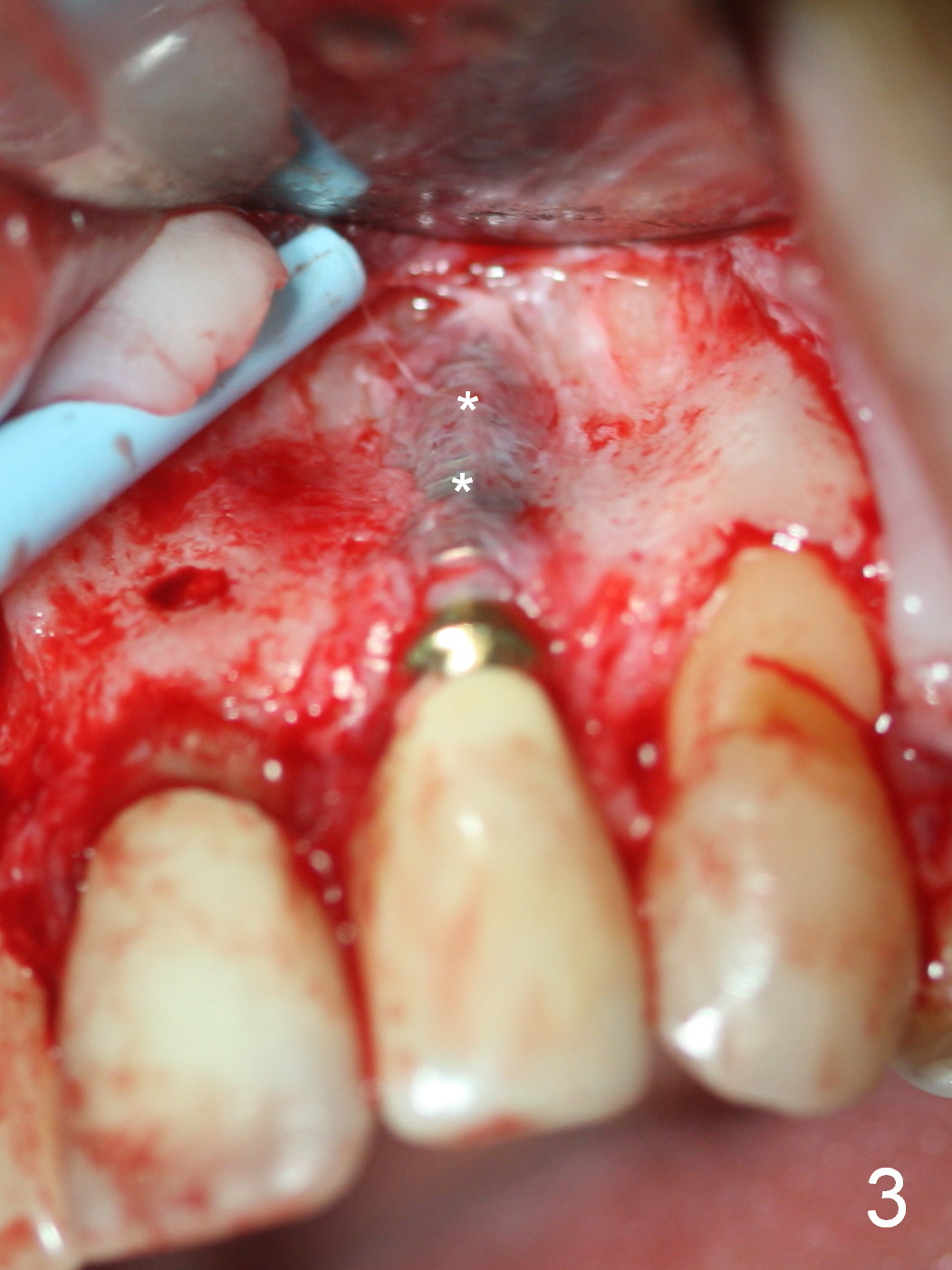
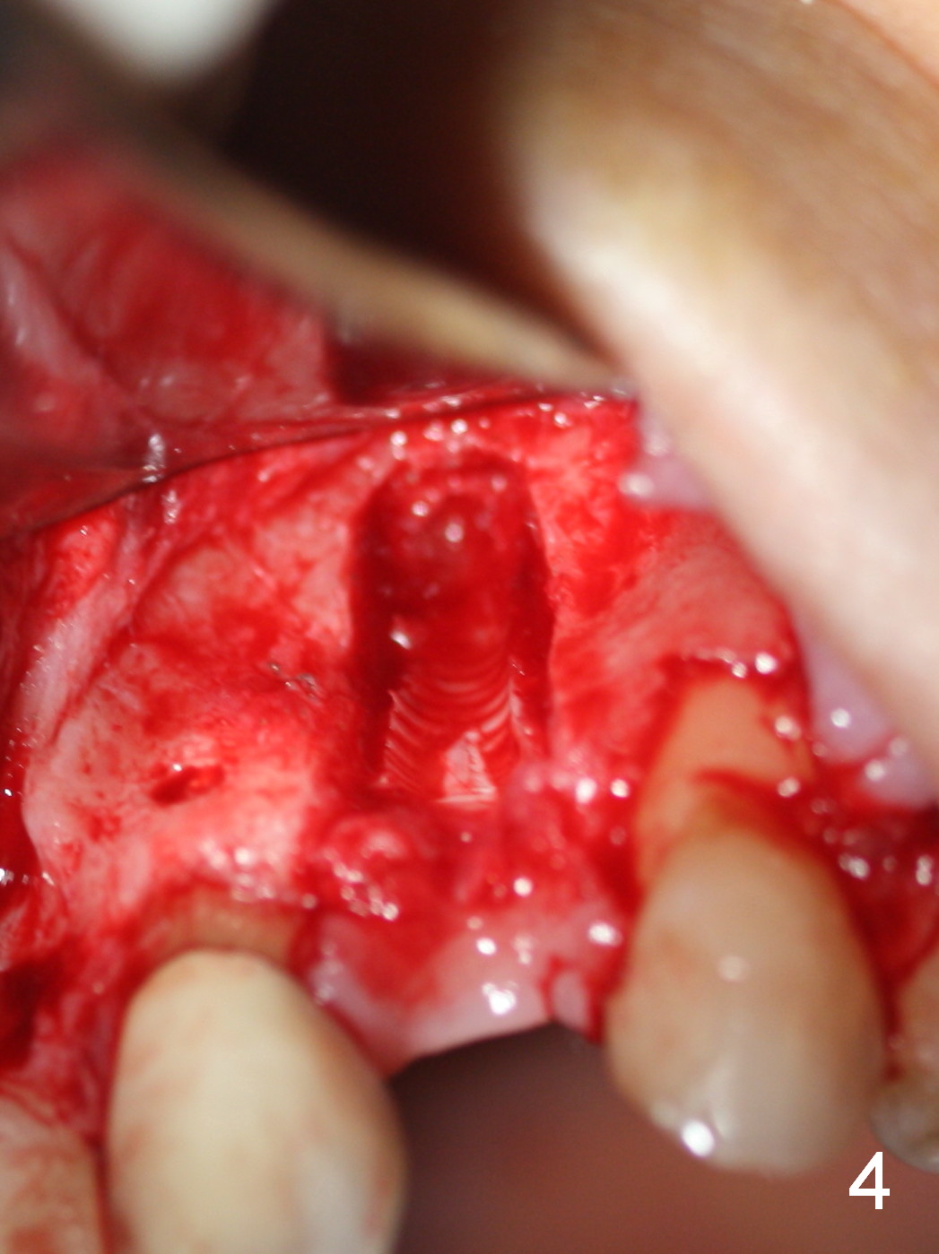
When the buccal flap is raised, the implant is found to be exposed buccally (Fig.3). When it is removed, the coronal portion of the palatal wall is thin (Fig.4). It appears that there is bone in the mid and apical thirds of the palatal wall, where an osteotomy is initiated using a 1.2 mm pilot drill.
Xin Wei, DDS, PhD, MS 1st edition 09/05/2015, last revision 01/19/2018